4GPV

Crystal structure of a putative cell adhesion protein (BACEGG_00536) from Bacteroides eggerthii DSM 20697 at 1.67 A resolution


Experimental Data Snapshot

  • Method: X-RAY DIFFRACTION
  • Resolution: 1.67 Å
  • R-Value Free: 0.189 
  • R-Value Work: 0.160 
  • R-Value Observed: 0.161 

wwPDB Validation   3D Report Full Report


This is version 1.4 of the entry. See complete history


Literature

A Distinct Type of Pilus from the Human Microbiome.

Xu, Q.Shoji, M.Shibata, S.Naito, M.Sato, K.Elsliger, M.A.Grant, J.C.Axelrod, H.L.Chiu, H.J.Farr, C.L.Jaroszewski, L.Knuth, M.W.Deacon, A.M.Godzik, A.Lesley, S.A.Curtis, M.A.Nakayama, K.Wilson, I.A.

(2016) Cell 165: 690-703

  • DOI: https://doi.org/10.1016/j.cell.2016.03.016
  • Primary Citation of Related Structures:  
    3LIU, 3PAY, 3R4R, 3SY6, 3T2L, 3UFI, 3UP6, 4DGU, 4EPS, 4GPV, 4H40, 4JG5, 4JRF, 4K4K, 4Q98, 4QB7, 4QDG, 4RDB, 5CAG

  • PubMed Abstract: 

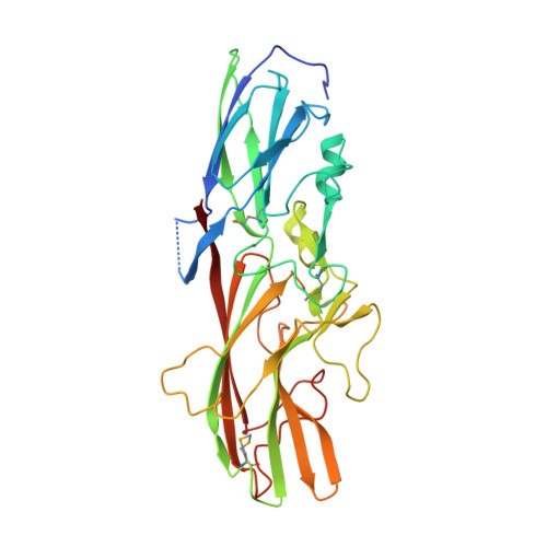
    Pili are proteinaceous polymers of linked pilins that protrude from the cell surface of many bacteria and often mediate adherence and virulence. We investigated a set of 20 Bacteroidia pilins from the human microbiome whose structures and mechanism of assembly were unknown. Crystal structures and biochemical data revealed a diverse protein superfamily with a common Greek-key β sandwich fold with two transthyretin-like repeats that polymerize into a pilus through a strand-exchange mechanism. The assembly mechanism of the central, structural pilins involves proteinase-assisted removal of their N-terminal β strand, creating an extended hydrophobic groove that binds the C-terminal donor strands of the incoming pilin. Accessory pilins at the tip and base have unique structural features specific to their location, allowing initiation or termination of the assembly. The Bacteroidia pilus, therefore, has a biogenesis mechanism that is distinct from other known pili and likely represents a different type of bacterial pilus.


  • Organizational Affiliation

    Joint Center for Structural Genomics, http://www.jcsg.org; SLAC National Accelerator Laboratory, Stanford Synchrotron Radiation Lightsource, Menlo Park, CA 94025, USA.


Macromolecules
Find similar proteins by:  (by identity cutoff)  |  3D Structure
Entity ID: 1
MoleculeChains Sequence LengthOrganismDetailsImage
putative cell adhesion protein
A, B
348Bacteroides eggerthii DSM 20697Mutation(s): 0 
Gene Names: BACEGG_00536
Entity Groups  
Sequence Clusters30% Identity50% Identity70% Identity90% Identity95% Identity100% Identity
Sequence Annotations
Expand
  • Reference Sequence
Small Molecules
Ligands 1 Unique
IDChains Name / Formula / InChI Key2D Diagram3D Interactions
TRS
Query on TRS

Download Ideal Coordinates CCD File 
C [auth A],
D [auth B]
2-AMINO-2-HYDROXYMETHYL-PROPANE-1,3-DIOL
C4 H12 N O3
LENZDBCJOHFCAS-UHFFFAOYSA-O
Modified Residues  1 Unique
IDChains TypeFormula2D DiagramParent
MSE
Query on MSE
A, B
L-PEPTIDE LINKINGC5 H11 N O2 SeMET
Experimental Data & Validation

Experimental Data

  • Method: X-RAY DIFFRACTION
  • Resolution: 1.67 Å
  • R-Value Free: 0.189 
  • R-Value Work: 0.160 
  • R-Value Observed: 0.161 
  • Space Group: P 1
  • Diffraction Data: https://doi.org/10.18430/M34GPV
Unit Cell:
Length ( Å )Angle ( ˚ )
a = 46.06α = 109.61
b = 61.488β = 103.02
c = 64.112γ = 90.9
Software Package:
Software NamePurpose
MolProbitymodel building
PDB_EXTRACTdata extraction
SHELXphasing
SHARPphasing
XSCALEdata scaling
BUSTER-TNTrefinement
XDSdata reduction
SHELXDphasing
BUSTERrefinement

Structure Validation

View Full Validation Report


Entry History 

Deposition Data

Revision History  (Full details and data files)

  • Version 1.0: 2012-09-19
    Type: Initial release
  • Version 1.1: 2017-11-15
    Changes: Refinement description
  • Version 1.2: 2018-01-24
    Changes: Database references
  • Version 1.3: 2020-04-22
    Changes: Database references, Derived calculations
  • Version 1.4: 2023-02-01
    Changes: Database references, Derived calculations