4GOS

Crystal structure of human B7-H4 IgV-like domain


Experimental Data Snapshot

  • Method: X-RAY DIFFRACTION
  • Resolution: 1.59 Å
  • R-Value Free: 0.192 
  • R-Value Work: 0.174 
  • R-Value Observed: 0.175 

wwPDB Validation   3D Report Full Report


This is version 2.0 of the entry. See complete history


Literature

Structure and cancer immunotherapy of the B7 family member B7x.

Jeon, H.Vigdorovich, V.Garrett-Thomson, S.C.Janakiram, M.Ramagopal, U.A.Abadi, Y.M.Lee, J.S.Scandiuzzi, L.Ohaegbulam, K.C.Chinai, J.M.Zhao, R.Yao, Y.Mao, Y.Sparano, J.A.Almo, S.C.Zang, X.

(2014) Cell Rep 9: 1089-1098

  • DOI: https://doi.org/10.1016/j.celrep.2014.09.053
  • Primary Citation of Related Structures:  
    4GOS

  • PubMed Abstract: 

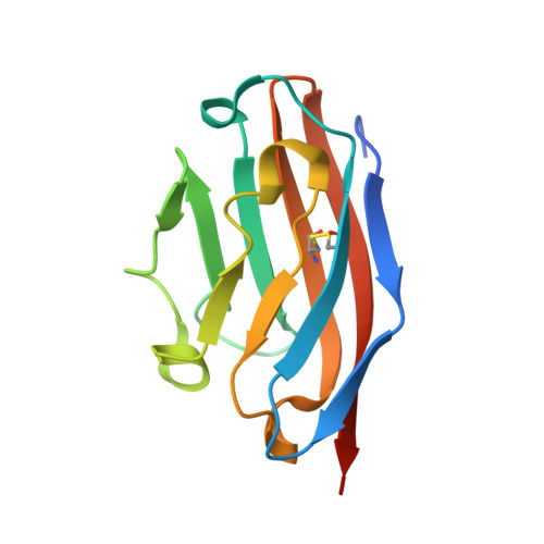
    B7x (B7-H4 or B7S1) is a member of the B7 family that can inhibit T cell function. B7x protein is absent in most normal human tissues and immune cells, but it is overexpressed in human cancers and often correlates with negative clinical outcome. The expression pattern and function of B7x suggest that it may be a potent immunosuppressive pathway in human cancers. Here, we determined the crystal structure of the human B7x immunoglobulin variable (IgV) domain at 1.59 Å resolution and mapped the epitopes recognized by monoclonal antibodies. We developed an in vivo system to screen therapeutic monoclonal antibodies against B7x and found that the clone 1H3 significantly inhibited growth of B7x-expressing tumors in vivo via multiple mechanisms. Furthermore, the surviving mice given 1H3 treatment were resistant to tumor rechallenge. Our data suggest that targeting B7x on tumors is a promising cancer immunotherapy and humanized 1H3 may be efficacious for immunotherapy of human cancers.


  • Organizational Affiliation

    Department of Microbiology and Immunology, Albert Einstein College of Medicine, Bronx, NY 10461, USA.


Macromolecules
Find similar proteins by:  (by identity cutoff)  |  3D Structure
Entity ID: 1
MoleculeChains Sequence LengthOrganismDetailsImage
V-set domain-containing T-cell activation inhibitor 1125Homo sapiensMutation(s): 0 
Gene Names: B7-H4B7H4B7S1B7xUNQ659/PRO1291VTCN1
UniProt & NIH Common Fund Data Resources
Find proteins for Q7Z7D3 (Homo sapiens)
Explore Q7Z7D3 
Go to UniProtKB:  Q7Z7D3
PHAROS:  Q7Z7D3
GTEx:  ENSG00000134258 
Entity Groups  
Sequence Clusters30% Identity50% Identity70% Identity90% Identity95% Identity100% Identity
UniProt GroupQ7Z7D3
Sequence Annotations
Expand
  • Reference Sequence
Oligosaccharides

Help

Entity ID: 2
MoleculeChains Length2D Diagram Glycosylation3D Interactions
alpha-D-mannopyranose-(1-3)-[alpha-D-mannopyranose-(1-6)]beta-D-mannopyranose-(1-4)-2-acetamido-2-deoxy-beta-D-glucopyranose-(1-4)-2-acetamido-2-deoxy-beta-D-glucopyranose
B
5N-Glycosylation
Glycosylation Resources
GlyTouCan:  G22768VO
GlyCosmos:  G22768VO
GlyGen:  G22768VO
Experimental Data & Validation

Experimental Data

  • Method: X-RAY DIFFRACTION
  • Resolution: 1.59 Å
  • R-Value Free: 0.192 
  • R-Value Work: 0.174 
  • R-Value Observed: 0.175 
  • Space Group: P 43 21 2
Unit Cell:
Length ( Å )Angle ( ˚ )
a = 46.5α = 90
b = 46.5β = 90
c = 115.77γ = 90
Software Package:
Software NamePurpose
SCALAdata scaling
SHELXphasing
REFMACrefinement
PDB_EXTRACTdata extraction
CBASSdata collection
MOSFLMdata reduction
SHELXDphasing

Structure Validation

View Full Validation Report


Entry History 

Revision History  (Full details and data files)

  • Version 1.0: 2012-09-12
    Type: Initial release
  • Version 1.1: 2016-11-16
    Changes: Database references
  • Version 2.0: 2020-07-29
    Type: Remediation
    Reason: Carbohydrate remediation
    Changes: Atomic model, Data collection, Database references, Derived calculations, Structure summary