4GB3

Human coxsackievirus B3 strain RD coat protein


Experimental Data Snapshot

  • Method: X-RAY DIFFRACTION
  • Resolution: 2.74 Å
  • R-Value Free: 0.339 
  • R-Value Work: 0.330 
  • R-Value Observed: 0.330 

wwPDB Validation   3D Report Full Report


Ligand Structure Quality Assessment 


This is version 1.2 of the entry. See complete history


Literature

The Crystal Structure of a Coxsackievirus B3-RD Variant and a Refined 9-Angstrom Cryo-Electron Microscopy Reconstruction of the Virus Complexed with Decay-Accelerating Factor (DAF) Provide a New Footprint of DAF on the Virus Surface.

Yoder, J.D.Cifuente, J.O.Pan, J.Bergelson, J.M.Hafenstein, S.

(2012) J Virol 86: 12571-12581

  • DOI: https://doi.org/10.1128/JVI.01592-12
  • Primary Citation of Related Structures:  
    3J24, 4GB3

  • PubMed Abstract: 

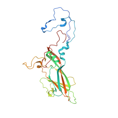
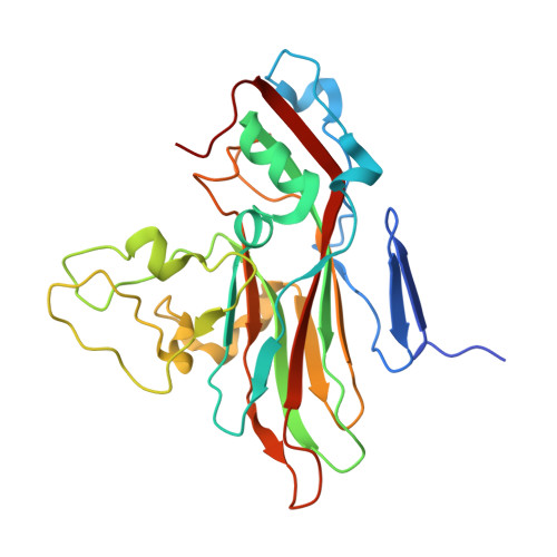
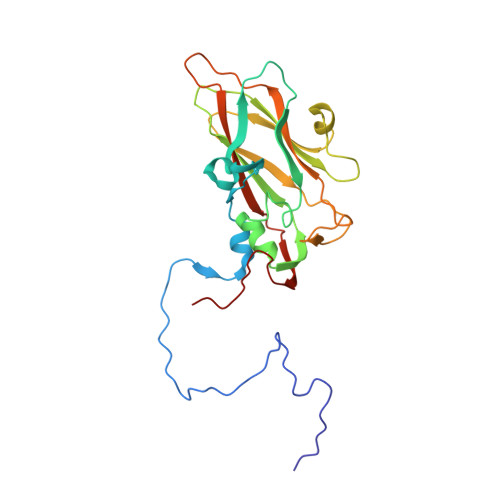
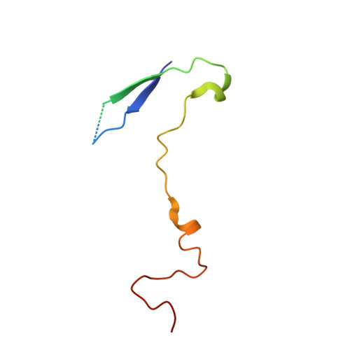
    The coxsackievirus-adenovirus receptor (CAR) and decay-accelerating factor (DAF) have been identified as cellular receptors for coxsackievirus B3 (CVB3). The first described DAF-binding isolate was obtained during passage of the prototype strain, Nancy, on rhabdomyosarcoma (RD) cells, which express DAF but very little CAR. Here, the structure of the resulting variant, CVB3-RD, has been solved by X-ray crystallography to 2.74 Å, and a cryo-electron microscopy reconstruction of CVB3-RD complexed with DAF has been refined to 9.0 Å. This new high-resolution structure permits us to correct an error in our previous view of DAF-virus interactions, providing a new footprint of DAF that bridges two adjacent protomers. The contact sites between the virus and DAF clearly encompass CVB3-RD residues recently shown to be required for binding to DAF; these residues interact with DAF short consensus repeat 2 (SCR2), which is known to be essential for virus binding. Based on the new structure, the mode of the DAF interaction with CVB3 differs significantly from the mode reported previously for DAF binding to echoviruses.


  • Organizational Affiliation

    The Pennsylvania State University College of Medicine, Hershey, Pennsylvania, USA.


Macromolecules
Find similar proteins by:  (by identity cutoff)  |  3D Structure
Entity ID: 1
MoleculeChains Sequence LengthOrganismDetailsImage
coat protein 1A [auth 1]281Coxsackievirus B3Mutation(s): 0 
UniProt
Find proteins for F8VA14 (Coxsackievirus B3)
Explore F8VA14 
Go to UniProtKB:  F8VA14
Entity Groups  
Sequence Clusters30% Identity50% Identity70% Identity90% Identity95% Identity100% Identity
UniProt GroupF8VA14
Sequence Annotations
Expand
  • Reference Sequence
Find similar proteins by:  (by identity cutoff)  |  3D Structure
Entity ID: 2
MoleculeChains Sequence LengthOrganismDetailsImage
coat protein 2B [auth 2]263Coxsackievirus B3Mutation(s): 0 
UniProt
Find proteins for F8VA14 (Coxsackievirus B3)
Explore F8VA14 
Go to UniProtKB:  F8VA14
Entity Groups  
Sequence Clusters30% Identity50% Identity70% Identity90% Identity95% Identity100% Identity
UniProt GroupF8VA14
Sequence Annotations
Expand
  • Reference Sequence
Find similar proteins by:  (by identity cutoff)  |  3D Structure
Entity ID: 3
MoleculeChains Sequence LengthOrganismDetailsImage
coat protein 3C [auth 3]238Coxsackievirus B3Mutation(s): 0 
UniProt
Find proteins for F8VA14 (Coxsackievirus B3)
Explore F8VA14 
Go to UniProtKB:  F8VA14
Entity Groups  
Sequence Clusters30% Identity50% Identity70% Identity90% Identity95% Identity100% Identity
UniProt GroupF8VA14
Sequence Annotations
Expand
  • Reference Sequence
Find similar proteins by:  (by identity cutoff)  |  3D Structure
Entity ID: 4
MoleculeChains Sequence LengthOrganismDetailsImage
coat protein 4D [auth 4]68Coxsackievirus B3Mutation(s): 0 
UniProt
Find proteins for F8VA14 (Coxsackievirus B3)
Explore F8VA14 
Go to UniProtKB:  F8VA14
Entity Groups  
Sequence Clusters30% Identity50% Identity70% Identity90% Identity95% Identity100% Identity
UniProt GroupF8VA14
Sequence Annotations
Expand
  • Reference Sequence
Small Molecules
Experimental Data & Validation

Experimental Data

  • Method: X-RAY DIFFRACTION
  • Resolution: 2.74 Å
  • R-Value Free: 0.339 
  • R-Value Work: 0.330 
  • R-Value Observed: 0.330 
  • Space Group: H 3 2
Unit Cell:
Length ( Å )Angle ( ˚ )
a = 296.638α = 90
b = 296.638β = 90
c = 813.215γ = 120
Software Package:
Software NamePurpose
ADSCdata collection
AMoREphasing
CNSrefinement
HKL-2000data reduction
HKL-2000data scaling

Structure Validation

View Full Validation Report


Ligand Structure Quality Assessment 


Entry History 

Deposition Data

Revision History  (Full details and data files)

  • Version 1.0: 2012-09-26
    Type: Initial release
  • Version 1.1: 2012-11-14
    Changes: Database references
  • Version 1.2: 2023-09-13
    Changes: Data collection, Database references, Derived calculations, Refinement description