4CZ9

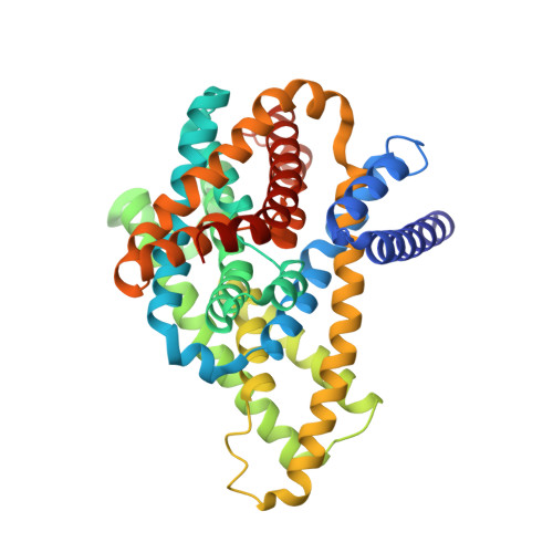
Structure of the sodium proton antiporter PaNhaP from Pyrococcus abyssii at pH 4.


Experimental Data Snapshot

  • Method: X-RAY DIFFRACTION
  • Resolution: 3.50 Å
  • R-Value Free: 0.263 
  • R-Value Work: 0.242 

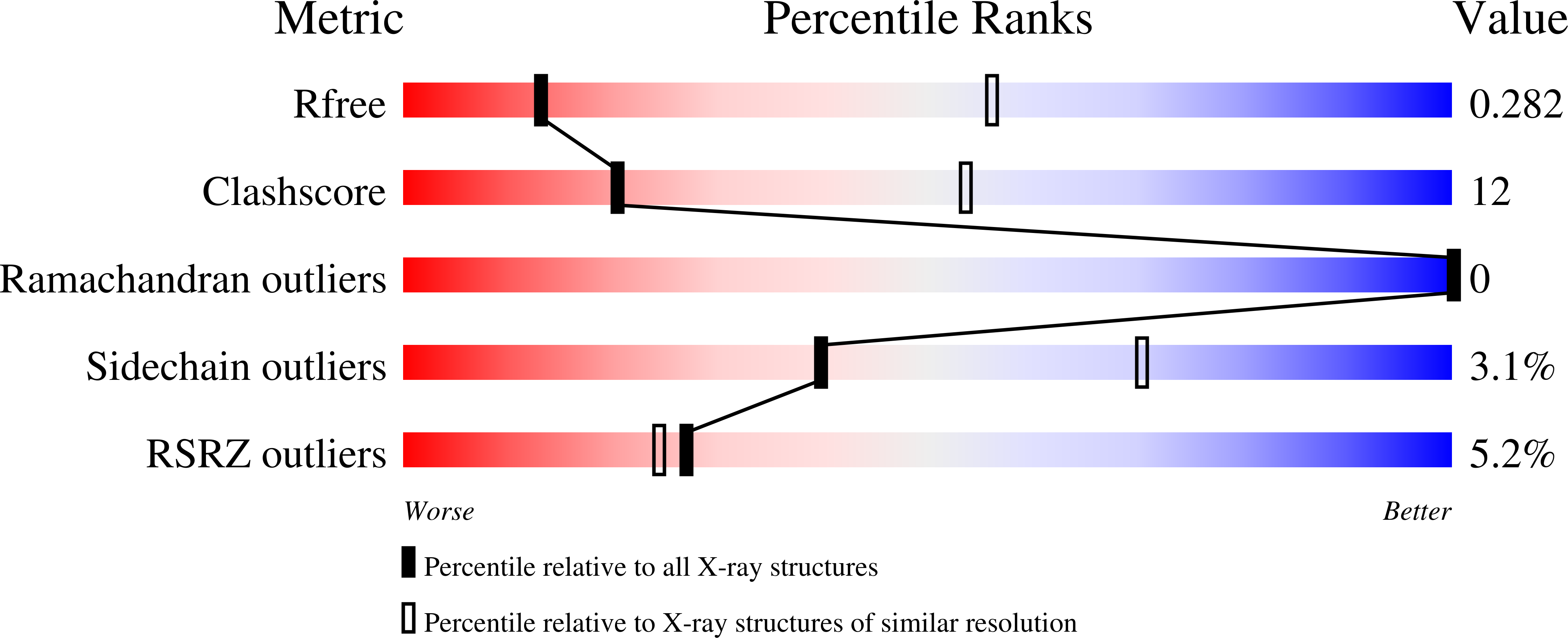
wwPDB Validation   3D Report Full Report


Ligand Structure Quality Assessment 


This is version 1.2 of the entry. See complete history


Literature

Structure and substrate ion binding in the sodium/proton antiporter PaNhaP.

Wohlert, D.Kuhlbrandt, W.Yildiz, O.

(2014) Elife 3: e03579-e03579

  • DOI: https://doi.org/10.7554/eLife.03579
  • Primary Citation of Related Structures:  
    4CZ8, 4CZ9, 4CZA

  • PubMed Abstract: 

    Sodium/proton antiporters maintain intracellular pH and sodium levels. Detailed structures of antiporters with bound substrate ions are essential for understanding how they work. We have resolved the substrate ion in the dimeric, electroneutral sodium/proton antiporter PaNhaP from Pyrococcus abyssi at 3.2 Å, and have determined its structure in two different conformations at pH 8 and pH 4. The ion is coordinated by three acidic sidechains, a water molecule, a serine and a main-chain carbonyl in the unwound stretch of trans-membrane helix 5 at the deepest point of a negatively charged cytoplasmic funnel. A second narrow polar channel may facilitate proton uptake from the cytoplasm. Transport activity of PaNhaP is cooperative at pH 6 but not at pH 5. Cooperativity is due to pH-dependent allosteric coupling of protomers through two histidines at the dimer interface. Combined with comprehensive transport studies, the structures of PaNhaP offer unique new insights into the transport mechanism of sodium/proton antiporters.


  • Organizational Affiliation

    Department of Structural Biology, Max Planck Institute of Biophysics, Frankfurt am Main, Germany.


Macromolecules
Find similar proteins by:  (by identity cutoff)  |  3D Structure
Entity ID: 1
MoleculeChains Sequence LengthOrganismDetailsImage
NA+/H+ ANTIPORTER, PUTATIVE
A, B
422Pyrococcus abyssi GE5Mutation(s): 0 
Membrane Entity: Yes 
UniProt
Find proteins for Q9UZ55 (Pyrococcus abyssi (strain GE5 / Orsay))
Explore Q9UZ55 
Go to UniProtKB:  Q9UZ55
Entity Groups  
Sequence Clusters30% Identity50% Identity70% Identity90% Identity95% Identity100% Identity
UniProt GroupQ9UZ55
Sequence Annotations
Expand
  • Reference Sequence
Small Molecules
Ligands 1 Unique
IDChains Name / Formula / InChI Key2D Diagram3D Interactions
OOC
Query on OOC

Download Ideal Coordinates CCD File 
C [auth B]octyl 4-O-beta-D-allopyranosyl-1-thio-beta-D-altropyranoside
C20 H38 O10 S
JHBBNAKIOKQRJS-AIIAIXEESA-N
Experimental Data & Validation

Experimental Data

  • Method: X-RAY DIFFRACTION
  • Resolution: 3.50 Å
  • R-Value Free: 0.263 
  • R-Value Work: 0.242 
  • Space Group: P 64
Unit Cell:
Length ( Å )Angle ( ˚ )
a = 109.566α = 90
b = 109.566β = 90
c = 209.626γ = 120
Software Package:
Software NamePurpose
PHENIXrefinement
XDSdata reduction
Aimlessdata scaling
PHASERphasing

Structure Validation

View Full Validation Report


Ligand Structure Quality Assessment 


Entry History 

Deposition Data

Revision History  (Full details and data files)

  • Version 1.0: 2014-12-17
    Type: Initial release
  • Version 1.1: 2019-02-27
    Changes: Data collection, Database references, Other
  • Version 1.2: 2023-12-20
    Changes: Data collection, Database references, Other, Refinement description