4B0R

Structure of the Deamidase-Depupylase Dop of the Prokaryotic Ubiquitin-like Modification Pathway


Experimental Data Snapshot

  • Method: X-RAY DIFFRACTION
  • Resolution: 2.60 Å
  • R-Value Free: 0.238 
  • R-Value Work: 0.194 
  • R-Value Observed: 0.196 

wwPDB Validation   3D Report Full Report


This is version 1.1 of the entry. See complete history


Literature

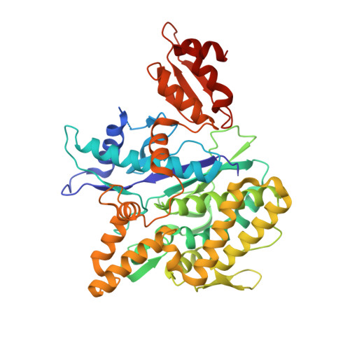
Structures of Pup ligase PafA and depupylase Dop from the prokaryotic ubiquitin-like modification pathway.

Ozcelik, D.Barandun, J.Schmitz, N.Sutter, M.Guth, E.Damberger, F.F.Allain, F.H.Ban, N.Weber-Ban, E.

(2012) Nat Commun 3: 1014-1014

  • DOI: https://doi.org/10.1038/ncomms2009
  • Primary Citation of Related Structures:  
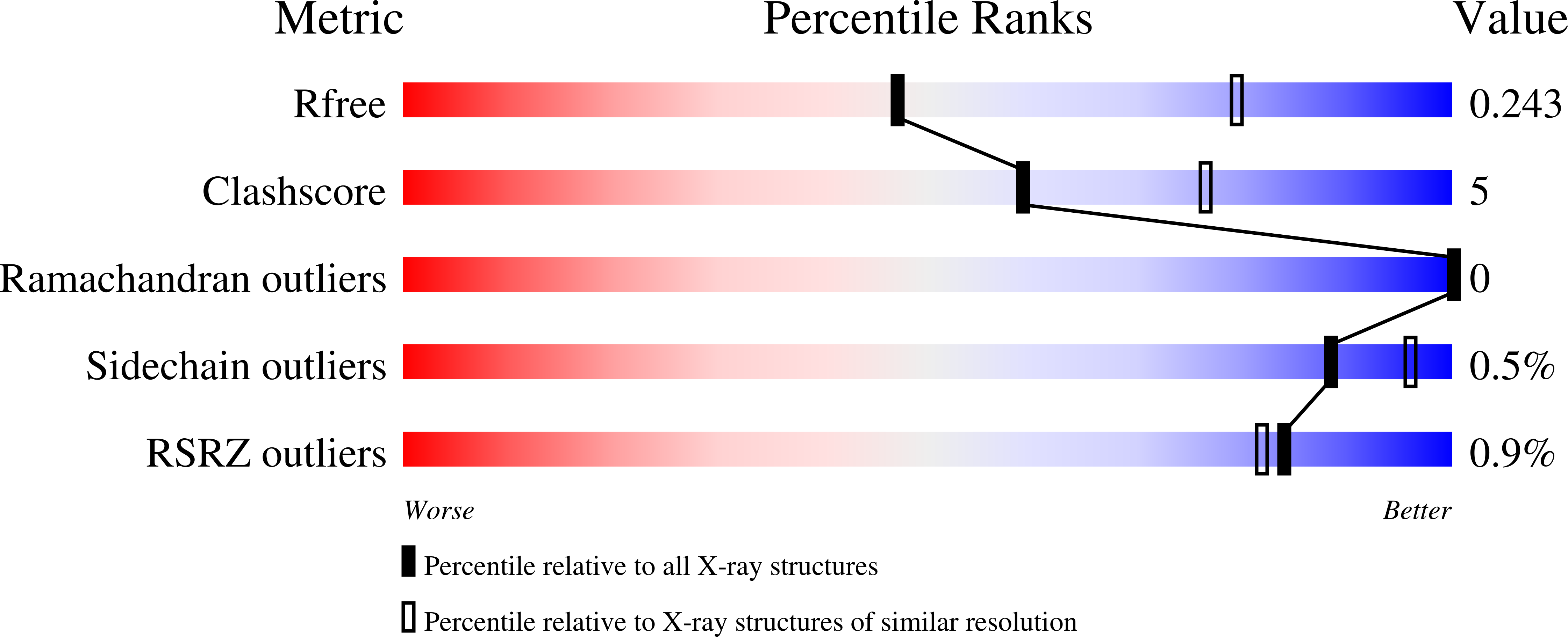
    4B0R, 4B0S, 4B0T

  • PubMed Abstract: 

    Pupylation is a posttranslational protein modification occurring in mycobacteria and other actinobacteria that is functionally analogous to ubiquitination. Here we report the crystal structures of the modification enzymes involved in this pathway, the prokaryotic ubiquitin-like protein (Pup) ligase PafA and the depupylase/deamidase Dop. Both feature a larger amino-terminal domain consisting of a central β-sheet packed against a cluster of helices, a fold characteristic for carboxylate-amine ligases, and a smaller C-terminal domain unique to PafA/Dop members. The active site is located on the concave surface of the β-sheet with the nucleotide bound in a deep pocket. A conserved groove leading into the active site could have a role in Pup-binding. Nuclear magnetic resonance and biochemical experiments determine the region of Pup that interacts with PafA and Dop. Structural data and mutational studies identify crucial residues for the catalysis of both enzymes.


  • Organizational Affiliation

    ETH Zurich, Institute of Molecular Biology & Biophysics, CH-8093, Switzerland.


Macromolecules
Find similar proteins by:  (by identity cutoff)  |  3D Structure
Entity ID: 1
MoleculeChains Sequence LengthOrganismDetailsImage
DEAMIDASE-DEPUPYLASE DOP506Acidothermus cellulolyticus 11BMutation(s): 0 
EC: 3.4 (PDB Primary Data), 3.5.1 (PDB Primary Data)
UniProt
Find proteins for A0LU48 (Acidothermus cellulolyticus (strain ATCC 43068 / DSM 8971 / 11B))
Explore A0LU48 
Go to UniProtKB:  A0LU48
Entity Groups  
Sequence Clusters30% Identity50% Identity70% Identity90% Identity95% Identity100% Identity
UniProt GroupA0LU48
Sequence Annotations
Expand
  • Reference Sequence
Experimental Data & Validation

Experimental Data

  • Method: X-RAY DIFFRACTION
  • Resolution: 2.60 Å
  • R-Value Free: 0.238 
  • R-Value Work: 0.194 
  • R-Value Observed: 0.196 
  • Space Group: P 21 21 21
Unit Cell:
Length ( Å )Angle ( ˚ )
a = 66.55α = 90
b = 71.47β = 90
c = 96.12γ = 90
Software Package:
Software NamePurpose
PHENIXrefinement
XDSdata reduction
XSCALEdata scaling
SHELXphasing

Structure Validation

View Full Validation Report


Entry History 

Deposition Data

Revision History  (Full details and data files)

  • Version 1.0: 2012-09-12
    Type: Initial release
  • Version 1.1: 2019-02-27
    Changes: Data collection, Database references, Experimental preparation, Other