4A3H

2',4' DINITROPHENYL-2-DEOXY-2-FLURO-B-D-CELLOBIOSIDE COMPLEX OF THE ENDOGLUCANASE CEL5A FROM BACILLUS AGARADHAERENS AT 1.6 A RESOLUTION


Experimental Data Snapshot

  • Method: X-RAY DIFFRACTION
  • Resolution: 1.65 Å
  • R-Value Free: 0.172 
  • R-Value Work: 0.142 

wwPDB Validation   3D Report Full Report


Ligand Structure Quality Assessment 


This is version 1.3 of the entry. See complete history


Literature

Snapshots along an enzymatic reaction coordinate: analysis of a retaining beta-glycoside hydrolase.

Davies, G.J.Mackenzie, L.Varrot, A.Dauter, M.Brzozowski, A.M.Schulein, M.Withers, S.G.

(1998) Biochemistry 37: 11707-11713

  • DOI: https://doi.org/10.1021/bi981315i
  • Primary Citation of Related Structures:  
    3A3H, 4A3H, 5A3H, 6A3H, 7A3H

  • PubMed Abstract: 

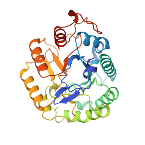
    The enzymatic hydrolysis of O-glycosidic linkages is one of the most diverse and widespread reactions in nature and involves a classic "textbook" enzyme mechanism. A multidisciplinary analysis of a beta-glycoside hydrolase, the Cel5A from Bacillus agaradhaerens, is presented in which the structures of each of the native, substrate, covalent-intermediate, and product complexes have been determined and their interconversions analyzed kinetically, providing unprecedented insights into the mechanism of this enzyme class. Substrate is bound in a distorted 1S3 skew-boat conformation, thereby presenting the anomeric carbon appropriately for nucleophilic attack as well as satisfying the stereoelectronic requirements for an incipient oxocarbenium ion. Leaving group departure results in the trapping of a covalent alpha-glycosyl-enzyme intermediate in which the sugar adopts an undistorted 4C1 conformation. Finally, hydrolysis of this intermediate yields a product complex in which the sugar is bound in a partially disordered mode, consistent with unfavorable interactions and low product affinity.


  • Organizational Affiliation

    Department of Chemistry, University of York, Heslington, U.K. davies@yorvic.york.ac.uk


Macromolecules
Find similar proteins by:  (by identity cutoff)  |  3D Structure
Entity ID: 1
MoleculeChains Sequence LengthOrganismDetailsImage
PROTEIN (ENDOGLUCANASE)303Salipaludibacillus agaradhaerensMutation(s): 0 
EC: 3.2.1.4
UniProt
Find proteins for O85465 (Salipaludibacillus agaradhaerens)
Explore O85465 
Go to UniProtKB:  O85465
Entity Groups  
Sequence Clusters30% Identity50% Identity70% Identity90% Identity95% Identity100% Identity
UniProt GroupO85465
Sequence Annotations
Expand
  • Reference Sequence
Small Molecules
Ligands 1 Unique
IDChains Name / Formula / InChI Key2D Diagram3D Interactions
DCB
Query on DCB

Download Ideal Coordinates CCD File 
B [auth A]2,4-DINITROPHENYL-2-DEOXY-2-FLUORO-BETA-D-CELLOBIOSIDE
C18 H23 F N2 O14
WFVCNOHOODVBQK-IUBYCILNSA-N
Experimental Data & Validation

Experimental Data

  • Method: X-RAY DIFFRACTION
  • Resolution: 1.65 Å
  • R-Value Free: 0.172 
  • R-Value Work: 0.142 
  • Space Group: P 21 21 21
Unit Cell:
Length ( Å )Angle ( ˚ )
a = 54.71α = 90
b = 69.57β = 90
c = 77.04γ = 90
Software Package:
Software NamePurpose
CCP4model building
REFMACrefinement
DENZOdata reduction
SCALEPACKdata scaling
CCP4phasing

Structure Validation

View Full Validation Report


Ligand Structure Quality Assessment 


Entry History 

Deposition Data

Revision History  (Full details and data files)

  • Version 1.0: 1999-07-23
    Type: Initial release
  • Version 1.1: 2007-10-16
    Changes: Version format compliance
  • Version 1.2: 2011-07-13
    Changes: Version format compliance
  • Version 1.3: 2023-12-27
    Changes: Data collection, Database references, Derived calculations