3ET6

The crystal structure of the catalytic domain of a eukaryotic guanylate cyclase


Experimental Data Snapshot

  • Method: X-RAY DIFFRACTION
  • Resolution: 2.55 Å
  • R-Value Free: 0.215 
  • R-Value Work: 0.172 
  • R-Value Observed: 0.174 

wwPDB Validation   3D Report Full Report


This is version 1.2 of the entry. See complete history


Literature

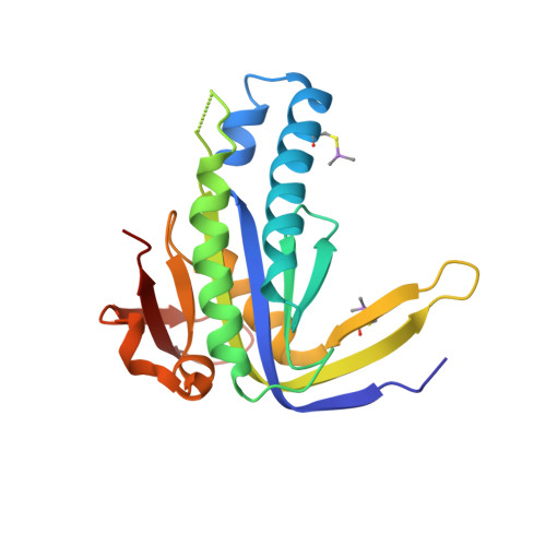
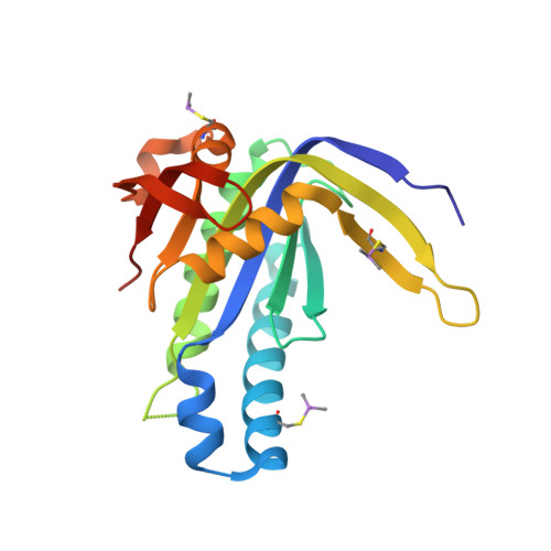
The crystal structure of the catalytic domain of a eukaryotic guanylate cyclase.

Winger, J.A.Derbyshire, E.R.Lamers, M.H.Marletta, M.A.Kuriyan, J.

(2008) BMC Struct Biol 8: 42-42

  • DOI: https://doi.org/10.1186/1472-6807-8-42
  • Primary Citation of Related Structures:  
    3ET6

  • PubMed Abstract: 

    Soluble guanylate cyclases generate cyclic GMP when bound to nitric oxide, thereby linking nitric oxide levels to the control of processes such as vascular homeostasis and neurotransmission. The guanylate cyclase catalytic module, for which no structure has been determined at present, is a class III nucleotide cyclase domain that is also found in mammalian membrane-bound guanylate and adenylate cyclases. We have determined the crystal structure of the catalytic domain of a soluble guanylate cyclase from the green algae Chlamydomonas reinhardtii at 2.55 A resolution, and show that it is a dimeric molecule. Comparison of the structure of the guanylate cyclase domain with the known structures of adenylate cyclases confirms the close similarity in architecture between these two enzymes, as expected from their sequence similarity. The comparison also suggests that the crystallized guanylate cyclase is in an inactive conformation, and the structure provides indications as to how activation might occur. We demonstrate that the two active sites in the dimer exhibit positive cooperativity, with a Hill coefficient of approximately 1.5. Positive cooperativity has also been observed in the homodimeric mammalian membrane-bound guanylate cyclases. The structure described here provides a reliable model for functional analysis of mammalian guanylate cyclases, which are closely related in sequence.


  • Organizational Affiliation

    Department of Molecular and Cell Biology, University of California, Berkeley, CA, USA. wingerj@berkeley.edu


Macromolecules
Find similar proteins by:  (by identity cutoff)  |  3D Structure
Entity ID: 1
MoleculeChains Sequence LengthOrganismDetailsImage
Soluble guanylyl cyclase beta190Chlamydomonas reinhardtiiMutation(s): 0 
Gene Names: CHLREDRAFT_187517CYG12
UniProt
Find proteins for Q5YLC2 (Chlamydomonas reinhardtii)
Explore Q5YLC2 
Go to UniProtKB:  Q5YLC2
Entity Groups  
Sequence Clusters30% Identity50% Identity70% Identity90% Identity95% Identity100% Identity
UniProt GroupQ5YLC2
Sequence Annotations
Expand
  • Reference Sequence
Find similar proteins by:  (by identity cutoff)  |  3D Structure
Entity ID: 2
MoleculeChains Sequence LengthOrganismDetailsImage
Soluble guanylyl cyclase beta190Chlamydomonas reinhardtiiMutation(s): 0 
Gene Names: CHLREDRAFT_187517CYG12
UniProt
Find proteins for Q5YLC2 (Chlamydomonas reinhardtii)
Explore Q5YLC2 
Go to UniProtKB:  Q5YLC2
Entity Groups  
Sequence Clusters30% Identity50% Identity70% Identity90% Identity95% Identity100% Identity
UniProt GroupQ5YLC2
Sequence Annotations
Expand
  • Reference Sequence
Experimental Data & Validation

Experimental Data

  • Method: X-RAY DIFFRACTION
  • Resolution: 2.55 Å
  • R-Value Free: 0.215 
  • R-Value Work: 0.172 
  • R-Value Observed: 0.174 
  • Space Group: P 32 2 1
Unit Cell:
Length ( Å )Angle ( ˚ )
a = 123.678α = 90
b = 123.678β = 90
c = 62.822γ = 120
Software Package:
Software NamePurpose
MOSFLMdata reduction
SCALAdata scaling
PHASERphasing
PHENIXrefinement
PDB_EXTRACTdata extraction
BOSdata collection

Structure Validation

View Full Validation Report


Entry History 

Deposition Data

Revision History  (Full details and data files)

  • Version 1.0: 2008-10-14
    Type: Initial release
  • Version 1.1: 2011-07-13
    Changes: Version format compliance
  • Version 1.2: 2023-09-06
    Changes: Data collection, Database references, Derived calculations, Refinement description