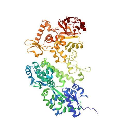
Crystal structure of the C-terminal globular domain of the third paralog of the Archaeoglobus fulgidus oligosaccharyltransferases
Matsumoto, S., Shimada, A., Kohda, D.(2013) BMC Struct Biol 13: 11-11
- PubMed: 23815857
- DOI: https://doi.org/10.1186/1472-6807-13-11
- Primary Citation of Related Structures:
3WAI - PubMed Abstract:
Protein N-glycosylation occurs in the three domains of life. Oligosaccharyltransferase (OST) transfers an oligosaccharide chain to the asparagine residue in the N-glycosylation sequons. The catalytic subunits of the OST enzyme are STT3 in eukaryotes, AglB in archaea and PglB in eubacteria. The genome of a hyperthermophilic archaeon, Archaeoglobus fulgidus, encodes three paralogous AglB proteins. We previously solved the crystal structures of the C-terminal globular domains of two paralogs, AglB-Short 1 and AglB-Short 2. We determined the crystal structure of the C-terminal globular domain of the third AglB paralog, AglB-Long, at 1.9 Å resolutions. The crystallization of the fusion protein with maltose binding protein (MBP) afforded high quality protein crystals. Two MBP-AglB-L molecules formed a swapped dimer in the crystal. Since the fusion protein behaved as a monomer upon gel filtration, we reconstituted the monomer structure from the swapped dimer by exchanging the swapped segments. The C-terminal domain of A. fulgidus AglB-L includes a structural unit common to AglB-S1 and AglB-S2. This structural unit contains the evolutionally conserved WWDYG and DK motifs. The present structure revealed that A. fulgidus AglB-L contained a variant type of the DK motif with a short insertion, and confirmed that the second signature residue, Lys, of the DK motif participates in the formation of a pocket that binds to the serine and threonine residues at the +2 position of the N-glycosylation sequon. The structure of A. fulgidus AglB-L, together with the two previously solved structures of AglB-S1 and AglB-S2, provides a complete overview of the three AglB paralogs encoded in the A. fulgidus genome. All three AglBs contain a variant type of the DK motif. This finding supports a previously proposed rule: The STT3/AglB/PglB paralogs in one organism always contain the same type of Ser/Thr-binding pocket. The present structure will be useful as a search model for molecular replacement in the structural determination of the full-length A. fulgidus AglB-L.