3VMA

Crystal Structure of the Full-Length Transglycosylase PBP1b from Escherichia coli


Experimental Data Snapshot

  • Method: X-RAY DIFFRACTION
  • Resolution: 2.16 Å
  • R-Value Free: 0.250 
  • R-Value Work: 0.212 
  • R-Value Observed: 0.214 

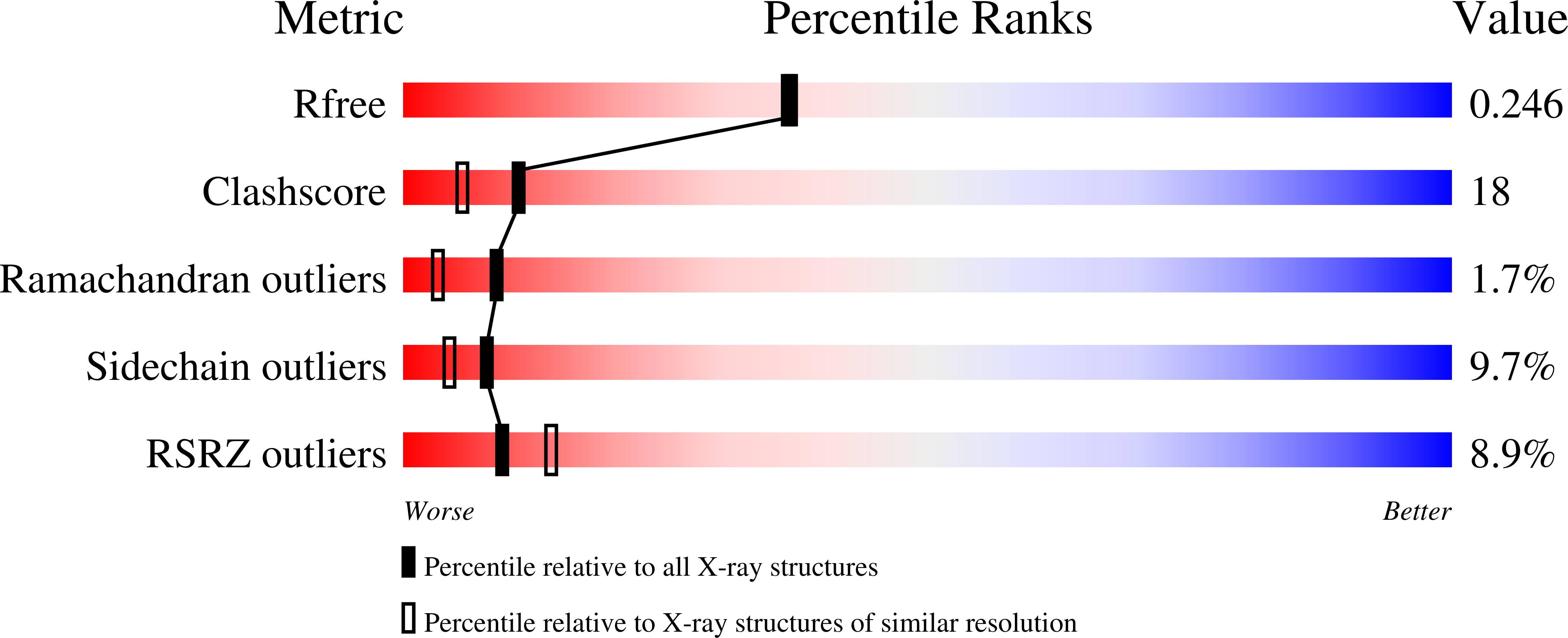
wwPDB Validation   3D Report Full Report


Ligand Structure Quality Assessment 


This is version 1.1 of the entry. See complete history


Literature

Crystal structure of the membrane-bound bifunctional transglycosylase PBP1b from Escherichia coli.

Sung, M.T.Lai, Y.T.Huang, C.Y.Chou, L.Y.Shih, H.W.Cheng, W.C.Wong, C.H.Ma, C.

(2009) Proc Natl Acad Sci U S A 106: 8824-8829

  • DOI: https://doi.org/10.1073/pnas.0904030106
  • Primary Citation of Related Structures:  
    3FWL, 3VMA

  • PubMed Abstract: 

    Drug-resistant bacteria have caused serious medical problems in recent years, and the need for new antibacterial agents is undisputed. Transglycosylase, a multidomain membrane protein essential for cell wall synthesis, is an excellent target for the development of new antibiotics. Here, we determined the X-ray crystal structure of the bifunctional transglycosylase penicillin-binding protein 1b (PBP1b) from Escherichia coli in complex with its inhibitor moenomycin to 2.16-A resolution. In addition to the transglycosylase and transpeptidase domains, our structure provides a complete visualization of this important antibacterial target, and reveals a domain for protein-protein interaction and a transmembrane helix domain essential for substrate binding, enzymatic activity, and membrane orientation.


  • Organizational Affiliation

    Genomics Research Center, Academia Sinica, 128 Academia Road, Section 2, Taipei 115, Taiwan.


Macromolecules
Find similar proteins by:  (by identity cutoff)  |  3D Structure
Entity ID: 1
MoleculeChains Sequence LengthOrganismDetailsImage
Penicillin-binding protein 1B768Escherichia coli K-12Mutation(s): 0 
Gene Names: b0149JW0145mrcBpbpFponB
EC: 2.4.1.129 (PDB Primary Data), 3.4 (PDB Primary Data)
Membrane Entity: Yes 
UniProt
Find proteins for P02919 (Escherichia coli (strain K12))
Explore P02919 
Go to UniProtKB:  P02919
Entity Groups  
Sequence Clusters30% Identity50% Identity70% Identity90% Identity95% Identity100% Identity
UniProt GroupP02919
Sequence Annotations
Expand
  • Reference Sequence
Small Molecules
Ligands 1 Unique
IDChains Name / Formula / InChI Key2D Diagram3D Interactions
M0E
Query on M0E

Download Ideal Coordinates CCD File 
B [auth A]MOENOMYCIN
C69 H106 N5 O34 P
NXPRJQIAIORCGO-ZLPAOQQDSA-N
Experimental Data & Validation

Experimental Data

  • Method: X-RAY DIFFRACTION
  • Resolution: 2.16 Å
  • R-Value Free: 0.250 
  • R-Value Work: 0.212 
  • R-Value Observed: 0.214 
  • Space Group: P 21 21 2
Unit Cell:
Length ( Å )Angle ( ˚ )
a = 62.9α = 90
b = 289.53β = 90
c = 62.78γ = 90
Software Package:
Software NamePurpose
HKL-2000data collection
SOLVEphasing
PHENIXrefinement
HKL-2000data reduction
HKL-2000data scaling

Structure Validation

View Full Validation Report


Ligand Structure Quality Assessment 


Entry History 

Deposition Data

Revision History  (Full details and data files)

  • Version 1.0: 2012-03-14
    Type: Initial release
  • Version 1.1: 2023-11-08
    Changes: Data collection, Database references, Derived calculations, Refinement description