3VID

Crystal structure of human VEGFR2 kinase domain with Compound A.


Experimental Data Snapshot

  • Method: X-RAY DIFFRACTION
  • Resolution: 2.30 Å
  • R-Value Free: 0.281 
  • R-Value Work: 0.245 
  • R-Value Observed: 0.247 

wwPDB Validation   3D Report Full Report


Ligand Structure Quality Assessment 


This is version 1.2 of the entry. See complete history


Literature

A Back-to-Front Fragment-Based Drug Design Search Strategy Targeting the DFG-Out Pocket of Protein Tyrosine Kinases.

Iwata, H.Oki, H.Okada, K.Takagi, T.Tawada, M.Miyazaki, Y.Imamura, S.Hori, A.Lawson, J.D.Hixon, M.S.Kimura, H.Miki, H.

(2012) ACS Med Chem Lett 3: 342-346

  • DOI: https://doi.org/10.1021/ml3000403
  • Primary Citation of Related Structures:  
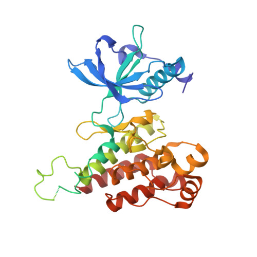
    3VHK, 3VID

  • PubMed Abstract: 

    We present a straightforward process for the discovery of novel back pocket-binding fragment molecules against protein tyrosine kinases. The approach begins by screening against the nonphosphorylated target kinase with subsequent counterscreening of hits against the phosphorylated enzyme. Back pocket-binding fragments are inactive against the phosphorylated kinase. Fragment molecules are of insufficient size to span both regions of the ATP binding pocket; thus, the outcome is binary (back pocket-binding or hinge-binding). Next, fragments with the appropriate binding profile are assayed in combination with a known hinge-binding fragment and subsequently with a known back pocket-binding fragment. Confirmation of back pocket-binding by Yonetani-Theorell plot analysis progresses candidate fragments to crystallization trials. The method is exemplified by a fragment screening campaign against vascular endothelial growth factor receptor 2, and a novel back pocket-binding fragment is presented.


  • Organizational Affiliation

    Discovery Research Laboratories, Oncology Drug Discovery Unit, Pharmaceutical Research Division, Takeda Pharmaceutical Company Ltd. , 26-1, Muraoka-Higashi 2-chome, Fujisawa, Kanagawa 251-8555, Japan.


Macromolecules
Find similar proteins by:  (by identity cutoff)  |  3D Structure
Entity ID: 1
MoleculeChains Sequence LengthOrganismDetailsImage
Vascular endothelial growth factor receptor 2356Homo sapiensMutation(s): 0 
Gene Names: KDRFLK1
EC: 2.7.10.1
UniProt & NIH Common Fund Data Resources
Find proteins for P35968 (Homo sapiens)
Explore P35968 
Go to UniProtKB:  P35968
PHAROS:  P35968
GTEx:  ENSG00000128052 
Entity Groups  
Sequence Clusters30% Identity50% Identity70% Identity90% Identity95% Identity100% Identity
UniProt GroupP35968
Sequence Annotations
Expand
  • Reference Sequence
Small Molecules
Ligands 1 Unique
IDChains Name / Formula / InChI Key2D Diagram3D Interactions
4TT
Query on 4TT

Download Ideal Coordinates CCD File 
B [auth A]4,5,6,11-tetrahydro-1H-pyrazolo[4',3':6,7]cyclohepta[1,2-b]indole
C14 H13 N3
HBWXPQZQCDQNND-UHFFFAOYSA-N
Binding Affinity Annotations 
IDSourceBinding Affinity
4TT PDBBind:  3VID IC50: 620 (nM) from 1 assay(s)
Experimental Data & Validation

Experimental Data

  • Method: X-RAY DIFFRACTION
  • Resolution: 2.30 Å
  • R-Value Free: 0.281 
  • R-Value Work: 0.245 
  • R-Value Observed: 0.247 
  • Space Group: P 21 21 21
Unit Cell:
Length ( Å )Angle ( ˚ )
a = 38.24α = 90
b = 94.62β = 90
c = 96.37γ = 90
Software Package:
Software NamePurpose
CrystalCleardata collection
AMoREphasing
REFMACrefinement
CrystalCleardata reduction
CrystalCleardata scaling

Structure Validation

View Full Validation Report


Ligand Structure Quality Assessment 


Entry History 

Deposition Data

Revision History  (Full details and data files)

  • Version 1.0: 2012-08-15
    Type: Initial release
  • Version 1.1: 2015-04-22
    Changes: Database references
  • Version 1.2: 2024-03-20
    Changes: Data collection, Database references, Derived calculations