3VD8

Crystal structure of human AIM2 PYD domain with MBP fusion


Experimental Data Snapshot

  • Method: X-RAY DIFFRACTION
  • Resolution: 2.07 Å
  • R-Value Free: 0.221 
  • R-Value Work: 0.182 
  • R-Value Observed: 0.184 

wwPDB Validation   3D Report Full Report


This is version 2.1 of the entry. See complete history


Literature

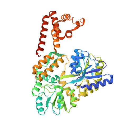
Structure of the Absent in Melanoma 2 (AIM2) Pyrin Domain Provides Insights into the Mechanisms of AIM2 Autoinhibition and Inflammasome Assembly.

Jin, T.Perry, A.Smith, P.Jiang, J.Xiao, T.S.

(2013) J Biol Chem 288: 13225-13235

  • DOI: https://doi.org/10.1074/jbc.M113.468033
  • Primary Citation of Related Structures:  
    3VD8

  • PubMed Abstract: 

    AIM2 binds dsDNA and associates with ASC through their PYDs to form an inflammasome. The AIM2 PYD structure illustrates distinct charge distribution and a unique hydrophobic patch. The AIM2 PYD may bind the ASC PYD and the AIM2 HIN domain through overlapping surface. These findings provide insights into the mechanisms of AIM2 autoinhibition and inflammasome assembly. Absent in melanoma 2 (AIM2) is a cytosolic double-stranded (dsDNA) sensor essential for innate immune responses against DNA viruses and bacteria such as Francisella and Listeria. Upon dsDNA engagement, the AIM2 amino-terminal pyrin domain (PYD) is responsible for downstream signaling to the adapter protein apoptosis-associated speck-like protein containing a caspase recruitment domain (ASC) through homotypic PYD-PYD interactions and the assembly of an inflammasome. Toward a better understanding of the AIM2 signaling mechanism, we determined the crystal structure of the human AIM2 PYD. The structure reveals a death domain fold with a short α3 helix that is buttressed by a highly conserved lysine residue at the α2 helix, which may stabilize the α3 helix for potential interaction with partner domains. The surface of the AIM2 PYD exhibits distinct charge distribution with highly acidic α1-α2 helices and highly basic α5-α6 helices. A prominent solvent-exposed hydrophobic patch formed by residues Phe-27 and Phe-28 at the α2 helix resembles a similar surface involved in the death effector domain homotypic interactions. Docking studies suggest that the AIM2 PYD may bind the AIM2 hematopoietic interferon-inducible nuclear (HIN) domain or ASC PYD using overlapping surface near the α2 helix. This may ensure that AIM2 interacts with the downstream adapter ASC only upon release of the autoinhibition by the dsDNA ligand. Our work thus unveils novel structural features of the AIM2 PYD and provides insights into the potential mechanisms of the PYD-HIN and PYD-PYD interactions important for AIM2 autoinhibition and inflammasome assembly.


  • Organizational Affiliation

    Structural Immunobiology Unit, Laboratory of Immunology, NIAID, National Institutes of Health, Bethesda, MD 20892-0430, USA.


Macromolecules
Find similar proteins by:  (by identity cutoff)  |  3D Structure
Entity ID: 1
MoleculeChains Sequence LengthOrganismDetailsImage
Maltose-binding periplasmic protein, Interferon-inducible protein AIM2489Escherichia coli O157:H7Homo sapiens
This entity is chimeric
Mutation(s): 5 
Gene Names: AIM2ECs5017malEMBPZ5632
UniProt & NIH Common Fund Data Resources
Find proteins for O14862 (Homo sapiens)
Explore O14862 
Go to UniProtKB:  O14862
PHAROS:  O14862
GTEx:  ENSG00000163568 
Find proteins for P0AEY0 (Escherichia coli O157:H7)
Explore P0AEY0 
Go to UniProtKB:  P0AEY0
Entity Groups  
Sequence Clusters30% Identity50% Identity70% Identity90% Identity95% Identity100% Identity
UniProt GroupsO14862P0AEY0
Sequence Annotations
Expand
  • Reference Sequence
Oligosaccharides

Help

Entity ID: 2
MoleculeChains Length2D Diagram Glycosylation3D Interactions
alpha-D-glucopyranose-(1-4)-alpha-D-glucopyranose-(1-4)-alpha-D-glucopyranose-(1-4)-alpha-D-glucopyranose
B
4N/A
Glycosylation Resources
GlyTouCan:  G87171PZ
GlyCosmos:  G87171PZ
GlyGen:  G87171PZ
Biologically Interesting Molecules (External Reference) 1 Unique
Experimental Data & Validation

Experimental Data

  • Method: X-RAY DIFFRACTION
  • Resolution: 2.07 Å
  • R-Value Free: 0.221 
  • R-Value Work: 0.182 
  • R-Value Observed: 0.184 
  • Space Group: P 21 21 21
Unit Cell:
Length ( Å )Angle ( ˚ )
a = 61.599α = 90
b = 91.934β = 90
c = 100.281γ = 90
Software Package:
Software NamePurpose
Blu-Icedata collection
PHASERphasing
PHENIXrefinement
HKL-2000data reduction
HKL-2000data scaling

Structure Validation

View Full Validation Report


Entry History 

Deposition Data

Revision History  (Full details and data files)

  • Version 1.0: 2013-01-16
    Type: Initial release
  • Version 1.1: 2013-02-13
    Changes: Database references, Structure summary
  • Version 1.2: 2013-06-05
    Changes: Database references
  • Version 1.3: 2017-08-02
    Changes: Refinement description, Source and taxonomy
  • Version 2.0: 2020-07-29
    Type: Remediation
    Reason: Carbohydrate remediation
    Changes: Atomic model, Data collection, Database references, Derived calculations, Non-polymer description, Structure summary
  • Version 2.1: 2023-09-13
    Changes: Data collection, Database references, Refinement description, Structure summary