3TM0

Crystal Structure of 3',5"-Aminoglycoside Phosphotransferase Type IIIa AMPPNP Butirosin A Complex


Experimental Data Snapshot

  • Method: X-RAY DIFFRACTION
  • Resolution: 2.10 Å
  • R-Value Free: 0.224 
  • R-Value Work: 0.179 
  • R-Value Observed: 0.184 

wwPDB Validation   3D Report Full Report


Ligand Structure Quality Assessment 


This is version 1.1 of the entry. See complete history


Literature

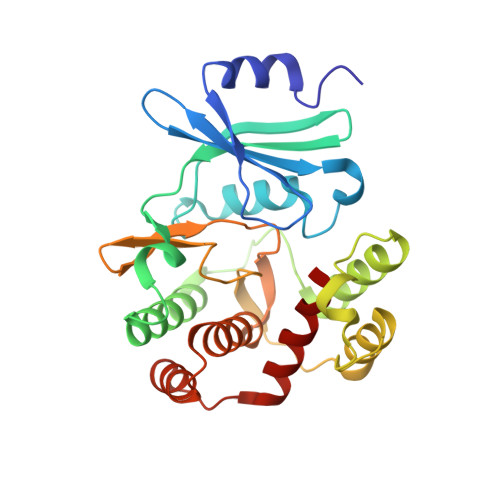
Structural basis of APH(3')-IIIa-mediated resistance to N1-substituted aminoglycoside antibiotics.

Fong, D.H.Berghuis, A.M.

(2009) Antimicrob Agents Chemother 53: 3049-3055

  • DOI: https://doi.org/10.1128/AAC.00062-09
  • Primary Citation of Related Structures:  
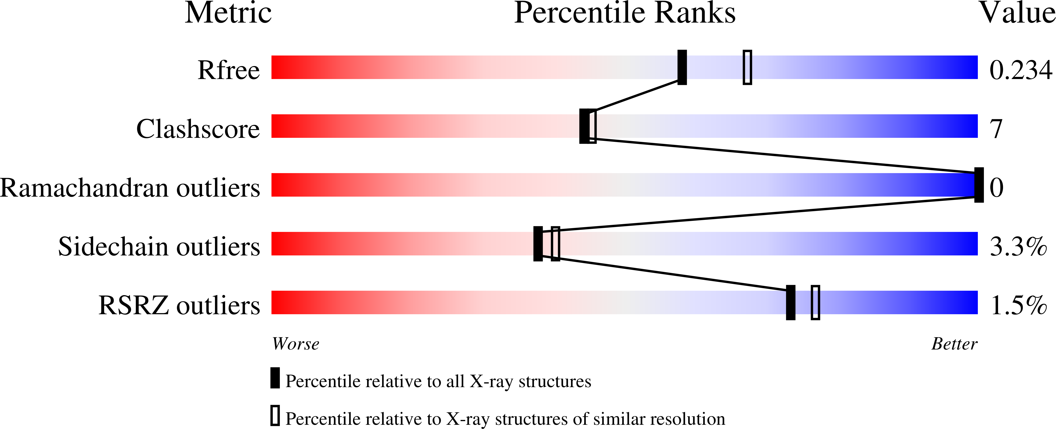
    3TM0

  • PubMed Abstract: 

    Butirosin is unique among the naturally occurring aminoglycosides, having a substituted amino group at position 1 (N1) of the 2-deoxystreptamine ring with an (S)-4-amino-2-hydroxybutyrate (AHB) group. While bacterial resistance to aminoglycosides can be ascribed chiefly to drug inactivation by plasmid-encoded aminoglycoside-modifying enzymes, the presence of an AHB group protects the aminoglycoside from binding to many resistance enzymes, and hence, the antibiotic retains its bactericidal properties. Consequently, several semisynthetic N1-substituted aminoglycosides, such as amikacin, isepamicin, and netilmicin, were developed. Unfortunately, butirosin, amikacin, and isepamicin are not resistant to inactivation by 3'-aminoglycoside O-phosphotransferase type IIIa [APH(3')-IIIa]. We report here the crystal structure of APH(3')-IIIa in complex with an ATP analog, AMPPNP [adenosine 5'-(beta,gamma-imido)triphosphate], and butirosin A to 2.4-A resolution. The structure shows that butirosin A binds to the enzyme in a manner analogous to other 4,5-disubstituted aminoglycosides, and the flexible antibiotic-binding loop is key to the accommodation of structurally diverse substrates. Based on the crystal structure, we have also constructed a model of APH(3')-IIIa in complex with amikacin, a commonly used semisynthetic N1-substituted 4,6-disubstituted aminoglycoside. Together, these results suggest a strategy to further derivatize the AHB group in order to generate new aminoglycoside derivatives that can elude inactivation by resistance enzymes while maintaining their ability to bind to the ribosomal A site.


  • Organizational Affiliation

    Department of Biochemistry, McGill University, 3649 Promenade Sir William Osler, Montreal, Quebec, Canada.


Macromolecules
Find similar proteins by:  (by identity cutoff)  |  3D Structure
Entity ID: 1
MoleculeChains Sequence LengthOrganismDetailsImage
Aminoglycoside 3'-phosphotransferase263Enterococcus faecalisMutation(s): 0 
Gene Names: aph(3')-iiiaaphA
EC: 2.7.1.95
UniProt
Find proteins for P0A3Y5 (Enterococcus faecalis)
Explore P0A3Y5 
Go to UniProtKB:  P0A3Y5
Entity Groups  
Sequence Clusters30% Identity50% Identity70% Identity90% Identity95% Identity100% Identity
UniProt GroupP0A3Y5
Sequence Annotations
Expand
  • Reference Sequence
Small Molecules
Ligands 3 Unique
IDChains Name / Formula / InChI Key2D Diagram3D Interactions
B31
Query on B31

Download Ideal Coordinates CCD File 
C [auth A](2S)-4-amino-N-[(1R,2S,3R,4R,5S)-5-amino-4-[(2,6-diamino-2,6-dideoxy-alpha-D-glucopyranosyl)oxy]-2-hydroxy-3-(beta-D-xylofuranosyloxy)cyclohexyl]-2-hydroxybutanamide
C21 H41 N5 O12
XEQLFNPSYWZPOW-SVRMBHBBSA-N
ANP
Query on ANP

Download Ideal Coordinates CCD File 
B [auth A]PHOSPHOAMINOPHOSPHONIC ACID-ADENYLATE ESTER
C10 H17 N6 O12 P3
PVKSNHVPLWYQGJ-KQYNXXCUSA-N
MG
Query on MG

Download Ideal Coordinates CCD File 
D [auth A],
E [auth A]
MAGNESIUM ION
Mg
JLVVSXFLKOJNIY-UHFFFAOYSA-N
Experimental Data & Validation

Experimental Data

  • Method: X-RAY DIFFRACTION
  • Resolution: 2.10 Å
  • R-Value Free: 0.224 
  • R-Value Work: 0.179 
  • R-Value Observed: 0.184 
  • Space Group: P 42 21 2
Unit Cell:
Length ( Å )Angle ( ˚ )
a = 80.112α = 90
b = 80.112β = 90
c = 110.6γ = 90
Software Package:
Software NamePurpose
CBASSdata collection
AMoREphasing
PHENIXrefinement
HKL-2000data reduction
HKL-2000data scaling

Structure Validation

View Full Validation Report


Ligand Structure Quality Assessment 


Entry History 

Deposition Data

Revision History  (Full details and data files)

  • Version 1.0: 2011-10-19
    Type: Initial release
  • Version 1.1: 2023-09-13
    Changes: Data collection, Database references, Derived calculations, Refinement description