3TJY

Structure of the Pto-binding domain of HopPmaL generated by limited chymotrypsin digestion


Experimental Data Snapshot

  • Method: X-RAY DIFFRACTION
  • Resolution: 1.70 Å
  • R-Value Free: 0.217 
  • R-Value Work: 0.188 
  • R-Value Observed: 0.189 

wwPDB Validation   3D Report Full Report


This is version 1.3 of the entry. See complete history


Literature

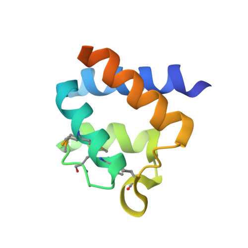
Structural analysis of HopPmaL reveals the presence of a second adaptor domain common to the HopAB family of Pseudomonas syringae type III effectors.

Singer, A.U.Wu, B.Yee, A.Houliston, S.Xu, X.Cui, H.Skarina, T.Garcia, M.Semesi, A.Arrowsmith, C.H.Savchenko, A.

(2012) Biochemistry 51: 1-3

  • DOI: https://doi.org/10.1021/bi2013883
  • Primary Citation of Related Structures:  
    2LF3, 2LF6, 3SVI, 3TJY

  • PubMed Abstract: 

    HopPmaL is a member of the HopAB family of type III effectors present in the phytopathogen Pseudomonas syringae. Using both X-ray crystallography and solution nuclear magnetic resonance, we demonstrate that HopPmaL contains two structurally homologous yet functionally distinct domains. The N-terminal domain corresponds to the previously described Pto-binding domain, while the previously uncharacterised C-terminal domain spans residues 308-385. While structurally similar, these domains do not share significant sequence similarity and most importantly demonstrate significant differences in key residues involved in host protein recognition, suggesting that each of them targets a different host protein.


  • Organizational Affiliation

    Department of Chemical Engineering and Applied Chemistry, Banting and Best Department of Medical Research, University of Toronto, Toronto, Ontario M5G 1L6, Canada.


Macromolecules
Find similar proteins by:  (by identity cutoff)  |  3D Structure
Entity ID: 1
MoleculeChains Sequence LengthOrganismDetailsImage
Effector protein hopAB394Pseudomonas syringae pv. maculicola str. ES4326Mutation(s): 0 
Gene Names: hopAB3hopPmaL
UniProt
Find proteins for Q8RP04 (Pseudomonas syringae pv. maculicola)
Explore Q8RP04 
Go to UniProtKB:  Q8RP04
Entity Groups  
Sequence Clusters30% Identity50% Identity70% Identity90% Identity95% Identity100% Identity
UniProt GroupQ8RP04
Sequence Annotations
Expand
  • Reference Sequence
Experimental Data & Validation

Experimental Data

  • Method: X-RAY DIFFRACTION
  • Resolution: 1.70 Å
  • R-Value Free: 0.217 
  • R-Value Work: 0.188 
  • R-Value Observed: 0.189 
  • Space Group: P 41 21 2
Unit Cell:
Length ( Å )Angle ( ˚ )
a = 57.427α = 90
b = 57.427β = 90
c = 54.8γ = 90
Software Package:
Software NamePurpose
PHENIXrefinement

Structure Validation

View Full Validation Report


Entry History 

Deposition Data

Revision History  (Full details and data files)

  • Version 1.0: 2011-09-14
    Type: Initial release
  • Version 1.1: 2012-01-11
    Changes: Database references
  • Version 1.2: 2012-02-22
    Changes: Structure summary
  • Version 1.3: 2013-01-09
    Changes: Database references