3SM1

The crystal structure of XMRV protease complexed with pepstatin A


Experimental Data Snapshot

  • Method: X-RAY DIFFRACTION
  • Resolution: 1.50 Å
  • R-Value Free: 0.196 
  • R-Value Work: 0.174 
  • R-Value Observed: 0.175 

wwPDB Validation   3D Report Full Report


This is version 1.3 of the entry. See complete history


Literature

Structural and biochemical characterization of the inhibitor complexes of xenotropic murine leukemia virus-related virus protease.

Li, M.Gustchina, A.Matuz, K.Tozser, J.Namwong, S.Goldfarb, N.E.Dunn, B.M.Wlodawer, A.

(2011) FEBS J 278: 4413-4424

  • DOI: https://doi.org/10.1111/j.1742-4658.2011.08364.x
  • Primary Citation of Related Structures:  
    3SLZ, 3SM1, 3SM2

  • PubMed Abstract: 

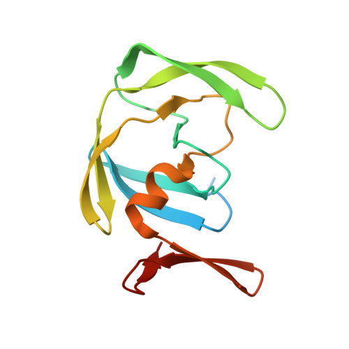
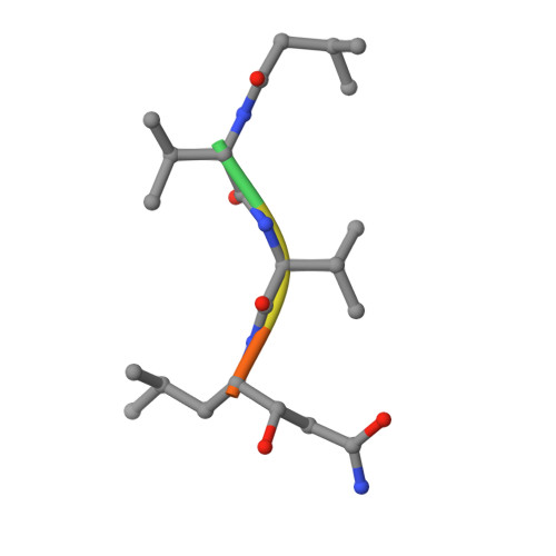
    Interactions between the protease (PR) encoded by the xenotropic murine leukemia virus-related virus and a number of potential inhibitors have been investigated by biochemical and structural techniques. It was observed that several inhibitors used clinically against HIV PR exhibit nanomolar or even subnanomolar values of K(i) , depending on the exact experimental conditions. Both TL-3, a universal inhibitor of retroviral PRs, and some inhibitors originally shown to inhibit plasmepsins were also quite potent, whereas inhibition by pepstatin A was considerably weaker. Crystal structures of the complexes of xenotropic murine leukemia virus-related virus PR with TL-3, amprenavir and pepstatin A were solved at high resolution and compared with the structures of complexes of these inhibitors with other retropepsins. Whereas TL-3 and amprenavir bound in a predictable manner, spanning the substrate-binding site of the enzyme, two molecules of pepstatin A bound simultaneously in an unprecedented manner, leaving the catalytic water molecule in place.


  • Organizational Affiliation

    Protein Structure Section, Macromolecular Crystallography Laboratory, National Cancer Institute, Frederick, MD 21702-1201, USA.


Macromolecules
Find similar proteins by:  (by identity cutoff)  |  3D Structure
Entity ID: 1
MoleculeChains Sequence LengthOrganismDetailsImage
gag-pro-pol polyprotein
A, B
132DG-75 Murine leukemia virusMutation(s): 0 
Gene Names: gag-pol
UniProt
Find proteins for Q9E7M1 (DG-75 Murine leukemia virus)
Explore Q9E7M1 
Go to UniProtKB:  Q9E7M1
Entity Groups  
Sequence Clusters30% Identity50% Identity70% Identity90% Identity95% Identity100% Identity
UniProt GroupQ9E7M1
Sequence Annotations
Expand
  • Reference Sequence

Find similar proteins by:  Sequence   |   3D Structure  

Entity ID: 2
MoleculeChains Sequence LengthOrganismDetailsImage
Pepstatin AC [auth J],
D [auth M]
6Streptomyces argenteolus subsp. toyonakensisMutation(s): 0 
Sequence Annotations
Expand
  • Reference Sequence
Biologically Interesting Molecules (External Reference) 1 Unique
Experimental Data & Validation

Experimental Data

  • Method: X-RAY DIFFRACTION
  • Resolution: 1.50 Å
  • R-Value Free: 0.196 
  • R-Value Work: 0.174 
  • R-Value Observed: 0.175 
  • Space Group: P 21 21 21
Unit Cell:
Length ( Å )Angle ( ˚ )
a = 46.399α = 90
b = 65.566β = 90
c = 69.775γ = 90
Software Package:
Software NamePurpose
APS-ID-22data collection
REFMACrefinement
HKL-3000data reduction
HKL-3000data scaling
REFMACphasing

Structure Validation

View Full Validation Report


Entry History 

Deposition Data

Revision History  (Full details and data files)

  • Version 1.0: 2011-10-12
    Type: Initial release
  • Version 1.1: 2012-04-04
    Changes: Database references
  • Version 1.2: 2012-12-12
    Changes: Other
  • Version 1.3: 2014-11-12
    Changes: Structure summary