


Crystal structure of methionine aminopeptidase 1b from Plasmodium Falciparum, PF10_0150
Wernimont, A.K., Artz, J.D., Crombet, L., Lew, J., Weadge, J., Arrowsmith, C.H., Edwards, A.M., Weigelt, J., Bountra, C., Hui, R., Hills, T.To be published.
Experimental Data Snapshot
wwPDB Validation 3D Report Full Report

Entity ID: 1 | |||||
|---|---|---|---|---|---|
| Molecule | Chains | Sequence Length | Organism | Details | Image |
| Methionine aminopeptidase | 368 | Plasmodium falciparum 3D7 | Mutation(s): 0 Gene Names: PF10_0150 EC: 3.4.11.18 | 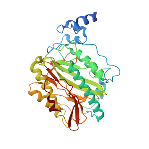 | |
UniProt | |||||
Find proteins for Q8IJP2 (Plasmodium falciparum (isolate 3D7)) Explore Q8IJP2 Go to UniProtKB: Q8IJP2 | |||||
Entity Groups | |||||
| Sequence Clusters | 30% Identity50% Identity70% Identity90% Identity95% Identity100% Identity | ||||
| UniProt Group | Q8IJP2 | ||||
Sequence AnnotationsExpand | |||||
| |||||
| Ligands 2 Unique | |||||
|---|---|---|---|---|---|
| ID | Chains | Name / Formula / InChI Key | 2D Diagram | 3D Interactions | |
| GOL Query on GOL | B [auth A], D [auth A] | GLYCEROL C3 H8 O3 PEDCQBHIVMGVHV-UHFFFAOYSA-N |  | ||
| FE Query on FE | C [auth A] | FE (III) ION Fe VTLYFUHAOXGGBS-UHFFFAOYSA-N |  | ||
| Length ( Å ) | Angle ( ˚ ) |
|---|---|
| a = 58.868 | α = 90 |
| b = 52.581 | β = 116.67 |
| c = 63.506 | γ = 90 |
| Software Name | Purpose |
|---|---|
| DENZO | data reduction |
| SCALEPACK | data scaling |
| REFMAC | refinement |
| PDB_EXTRACT | data extraction |
| JDirector | data collection |
| PHASER | phasing |
RCSB PDB (citation) is hosted by
RCSB PDB is a member of the

