3RFV

Crystal structure of Uronate dehydrogenase from Agrobacterium tumefaciens complexed with NADH and product


Experimental Data Snapshot

  • Method: X-RAY DIFFRACTION
  • Resolution: 2.10 Å
  • R-Value Free: 0.189 
  • R-Value Work: 0.157 
  • R-Value Observed: 0.159 

wwPDB Validation   3D Report Full Report


Ligand Structure Quality Assessment 


This is version 1.4 of the entry. See complete history


Literature

Crystal Structure of Uronate Dehydrogenase from Agrobacterium tumefaciens.

Parkkinen, T.Boer, H.Janis, J.Andberg, M.Penttila, M.Koivula, A.Rouvinen, J.

(2011) J Biol Chem 286: 27294-27300

  • DOI: https://doi.org/10.1074/jbc.M111.254854
  • Primary Citation of Related Structures:  
    3RFT, 3RFV, 3RFX

  • PubMed Abstract: 

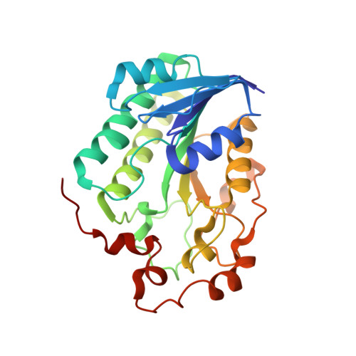
    Uronate dehydrogenase from Agrobacterium tumefaciens (AtUdh) belongs to the short-chain dehydrogenase/reductase superfamily and catalyzes the oxidation of D-galacturonic acid and D-glucuronic acid with NAD(+) as a cofactor. We have determined the crystal structures of an apo-form of AtUdh, a ternary form in complex with NADH and product (substrate-soaked structure), and an inactive Y136A mutant in complex with NAD(+). The crystal structures suggest AtUdh to be a homohexamer, which has also been observed to be the major form in solution. The monomer contains a Rossmann fold, essential for nucleotide binding and a common feature of the short-chain dehydrogenase/reductase family enzymes. The ternary complex structure reveals a product, D-galactaro-1,5-lactone, which is bound above the nicotinamide ring. This product rearranges in solution to D-galactaro-1,4-lactone as verified by mass spectrometry analysis, which agrees with our previous NMR study. The crystal structure of the mutant with the catalytic residue Tyr-136 substituted with alanine shows changes in the position of Ile-74 and Ser-75. This probably altered the binding of the nicotinamide end of NAD(+), which was not visible in the electron density map. The structures presented provide novel insights into cofactor and substrate binding and the reaction mechanism of AtUdh. This information can be applied to the design of efficient microbial conversion of D-galacturonic acid-based waste materials.


  • Organizational Affiliation

    Department of Chemistry, University of Eastern Finland, FI-80101 Joensuu, Finland.


Macromolecules
Find similar proteins by:  (by identity cutoff)  |  3D Structure
Entity ID: 1
MoleculeChains Sequence LengthOrganismDetailsImage
Uronate dehydrogenase
A, B, C
267Agrobacterium fabrum str. C58Mutation(s): 0 
Gene Names: udhAGR_L_3333Atu3143
EC: 1.1.1.203
UniProt
Find proteins for Q7CRQ0 (Agrobacterium fabrum (strain C58 / ATCC 33970))
Explore Q7CRQ0 
Go to UniProtKB:  Q7CRQ0
Entity Groups  
Sequence Clusters30% Identity50% Identity70% Identity90% Identity95% Identity100% Identity
UniProt GroupQ7CRQ0
Sequence Annotations
Expand
  • Reference Sequence
Small Molecules
Ligands 3 Unique
IDChains Name / Formula / InChI Key2D Diagram3D Interactions
NAI
Query on NAI

Download Ideal Coordinates CCD File 
D [auth A],
J [auth B],
N [auth C]
1,4-DIHYDRONICOTINAMIDE ADENINE DINUCLEOTIDE
C21 H29 N7 O14 P2
BOPGDPNILDQYTO-NNYOXOHSSA-N
15L
Query on 15L

Download Ideal Coordinates CCD File 
E [auth A],
K [auth B],
O [auth C]
D-galactaro-1,5-lactone
C6 H8 O7
YLKFQNUGXOLRNI-KXMYSMCESA-N
PO4
Query on PO4

Download Ideal Coordinates CCD File 
F [auth A]
G [auth A]
H [auth A]
I [auth A]
L [auth B]
F [auth A],
G [auth A],
H [auth A],
I [auth A],
L [auth B],
M [auth B],
P [auth C],
Q [auth C]
PHOSPHATE ION
O4 P
NBIIXXVUZAFLBC-UHFFFAOYSA-K
Experimental Data & Validation

Experimental Data

  • Method: X-RAY DIFFRACTION
  • Resolution: 2.10 Å
  • R-Value Free: 0.189 
  • R-Value Work: 0.157 
  • R-Value Observed: 0.159 
  • Space Group: P 62 2 2
Unit Cell:
Length ( Å )Angle ( ˚ )
a = 165.39α = 90
b = 165.39β = 90
c = 173.63γ = 120
Software Package:
Software NamePurpose
MOLREPphasing
PHENIXrefinement
XDSdata reduction
XSCALEdata scaling

Structure Validation

View Full Validation Report


Ligand Structure Quality Assessment 


Entry History 

Deposition Data

Revision History  (Full details and data files)

  • Version 1.0: 2011-06-15
    Type: Initial release
  • Version 1.1: 2011-07-13
    Changes: Version format compliance
  • Version 1.2: 2011-08-17
    Changes: Database references
  • Version 1.3: 2020-07-29
    Type: Remediation
    Reason: Carbohydrate remediation
    Changes: Data collection, Database references, Derived calculations, Structure summary
  • Version 1.4: 2023-09-13
    Changes: Data collection, Database references, Refinement description, Structure summary