3R93

Crystal structure of the chromo domain of M-phase phosphoprotein 8 bound to H3K9Me3 peptide


Experimental Data Snapshot

  • Method: X-RAY DIFFRACTION
  • Resolution: 2.06 Å
  • R-Value Free: 0.273 
  • R-Value Work: 0.220 
  • R-Value Observed: 0.222 

wwPDB Validation   3D Report Full Report


This is version 1.5 of the entry. See complete history


Literature

Structural basis for specific binding of human MPP8 chromodomain to histone H3 methylated at lysine 9.

Li, J.Li, Z.Ruan, J.Xu, C.Tong, Y.Pan, P.W.Tempel, W.Crombet, L.Min, J.Zang, J.

(2011) PLoS One 6: e25104-e25104

  • DOI: https://doi.org/10.1371/journal.pone.0025104
  • Primary Citation of Related Structures:  
    3LWE, 3R93

  • PubMed Abstract: 

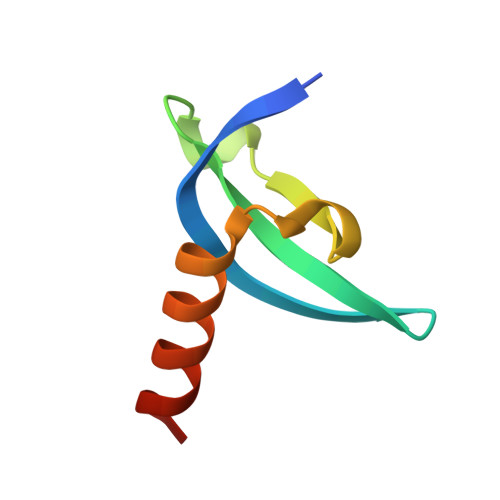
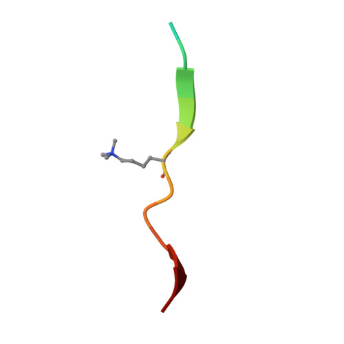
    M-phase phosphoprotein 8 (MPP8) was initially identified to be a component of the RanBPM-containing large protein complex, and has recently been shown to bind to methylated H3K9 both in vivo and in vitro. MPP8 binding to methylated H3K9 is suggested to recruit the H3K9 methyltransferases GLP and ESET, and DNA methyltransferase 3A to the promoter of the E-cadherin gene, mediating the E-cadherin gene silencing and promote tumor cell motility and invasion. MPP8 contains a chromodomain in its N-terminus, which is used to bind the methylated H3K9. Here, we reported the crystal structures of human MPP8 chromodomain alone and in complex with the trimethylated histone H3K9 peptide (residue 1-15). The complex structure unveils that the human MPP8 chromodomain binds methylated H3K9 through a conserved recognition mechanism, which was also observed in Drosophila HP1, a chromodomain containing protein that binds to methylated H3K9 as well. The structure also reveals that the human MPP8 chromodomain forms homodimer, which is mediated via an unexpected domain swapping interaction through two β strands from the two protomer subunits. Our findings reveal the molecular mechanism of selective binding of human MPP8 chromodomain to methylated histone H3K9. The observation of human MPP8 chromodomain in both solution and crystal lattice may provide clues to study MPP8-mediated gene regulation furthermore.


  • Organizational Affiliation

    Key Laboratory of Structural Biology, Chinese Academy of Sciences, and School of Life Sciences, University of Science and Technology of China, Hefei, Anhui, People's Republic of China.


Macromolecules
Find similar proteins by:  (by identity cutoff)  |  3D Structure
Entity ID: 1
MoleculeChains Sequence LengthOrganismDetailsImage
M-phase phosphoprotein 8
A, B, C, D
62Homo sapiensMutation(s): 0 
Gene Names: MPHOSPH8MPP8
UniProt & NIH Common Fund Data Resources
Find proteins for Q99549 (Homo sapiens)
Explore Q99549 
Go to UniProtKB:  Q99549
PHAROS:  Q99549
GTEx:  ENSG00000196199 
Entity Groups  
Sequence Clusters30% Identity50% Identity70% Identity90% Identity95% Identity100% Identity
UniProt GroupQ99549
Sequence Annotations
Expand
  • Reference Sequence

Find similar proteins by:  Sequence   |   3D Structure  

Entity ID: 2
MoleculeChains Sequence LengthOrganismDetailsImage
H3K9Me3 peptide
E, F, G, H
15Homo sapiensMutation(s): 0 
Gene Names: H3C15H3C14H3C13
UniProt & NIH Common Fund Data Resources
Find proteins for Q71DI3 (Homo sapiens)
Explore Q71DI3 
Go to UniProtKB:  Q71DI3
PHAROS:  Q71DI3
Entity Groups  
Sequence Clusters30% Identity50% Identity70% Identity90% Identity95% Identity100% Identity
UniProt GroupQ71DI3
Sequence Annotations
Expand
  • Reference Sequence
Small Molecules
Experimental Data & Validation

Experimental Data

  • Method: X-RAY DIFFRACTION
  • Resolution: 2.06 Å
  • R-Value Free: 0.273 
  • R-Value Work: 0.220 
  • R-Value Observed: 0.222 
  • Space Group: P 21 21 2
Unit Cell:
Length ( Å )Angle ( ˚ )
a = 71.148α = 90
b = 74.005β = 90
c = 72.609γ = 90
Software Package:
Software NamePurpose
DENZOdata reduction
SCALEPACKdata scaling
PHASERphasing
REFMACrefinement
PDB_EXTRACTdata extraction
HKL-2000data reduction
HKL-2000data scaling

Structure Validation

View Full Validation Report


Entry History 

Deposition Data

Revision History  (Full details and data files)

  • Version 1.0: 2011-04-06
    Type: Initial release
  • Version 1.1: 2011-07-13
    Changes: Version format compliance
  • Version 1.2: 2011-10-19
    Changes: Database references
  • Version 1.3: 2012-03-21
    Changes: Database references
  • Version 1.4: 2017-11-08
    Changes: Refinement description
  • Version 1.5: 2023-09-13
    Changes: Data collection, Database references, Derived calculations, Refinement description