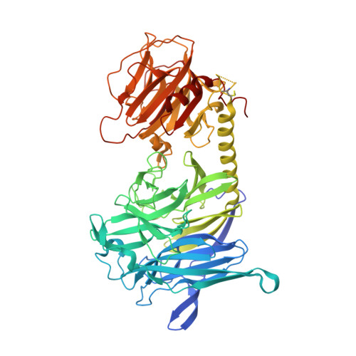
Crystal structure of an enzymatically inactive trans-sialidase-like lectin from Trypanosoma cruzi: the carbohydrate binding mechanism involves residual sialidase activity.
Oppezzo, P., Obal, G., Baraibar, M.A., Pritsch, O., Alzari, P.M., Buschiazzo, A.(2011) Biochim Biophys Acta 1814: 1154-1161
- PubMed: 21570497
- DOI: https://doi.org/10.1016/j.bbapap.2011.04.012
- Primary Citation of Related Structures:
3PJQ - PubMed Abstract:
Trans-sialidases are surface-located proteins in Trypanosoma cruzi that participate in key parasite-host interactions and parasite virulence. These proteins are encoded by a large multigenic family, with tandem-repeated and individual genes dispersed throughout the genome. While a large number of genes encode for catalytically active enzyme isoforms, many others display mutations that involve catalytic residues. The latter ultimately code for catalytically inactive proteins with very high similarity to their active paralogs. These inactive members have been shown to be lectins, able to bind sialic acid and galactose in vitro, although their cellular functions are yet to be fully established. We now report structural and biochemical evidence extending the current molecular understanding of these lectins. We have solved the crystal structure of one such catalytically inactive trans-sialidase-like protein, after soaking with a specific carbohydrate ligand, sialyl-α2,3-lactose. Instead of the expected trisaccharide, the binding pocket was observed occupied by α-lactose, strongly suggesting that the protein retains residual hydrolytic activity. This hypothesis was validated by enzyme kinetics assays, in comparison to fully active wild-type trans-sialidase. Surface plasmon resonance also confirmed that these trans-sialidase-like lectins are not only able to bind small oligosaccharides, but also sialylated glycoproteins, which is relevant in the physiologic scenario of parasite infection. Inactive trans-sialidase proteins appear thus to be β-methyl-galactosyl-specific lectins, evolved within an exo-sialidase scaffold, thus explaining why their lectin activity is triggered by the presence of terminal sialic acid.
Organizational Affiliation:
Institut Pasteur, Unité de Biochimie Structurale, Paris 75015, France.