3MAK

Crystal structure of Glutathione transferase dmGSTD1 from Drosophila melanogaster, in complex with glutathione


Experimental Data Snapshot

  • Method: X-RAY DIFFRACTION
  • Resolution: 1.80 Å
  • R-Value Free: 0.200 
  • R-Value Work: 0.160 
  • R-Value Observed: 0.162 

wwPDB Validation   3D Report Full Report


Ligand Structure Quality Assessment 


This is version 1.4 of the entry. See complete history


Literature

Structural evidence for conformational changes of Delta class glutathione transferases after ligand binding

Wongsantichon, J.Robinson, R.C.Ketterman, A.J.

(2012) Arch Biochem Biophys 521: 77-83

  • DOI: https://doi.org/10.1016/j.abb.2012.03.023
  • Primary Citation of Related Structures:  
    3F6F, 3G7I, 3GH6, 3MAK

  • PubMed Abstract: 

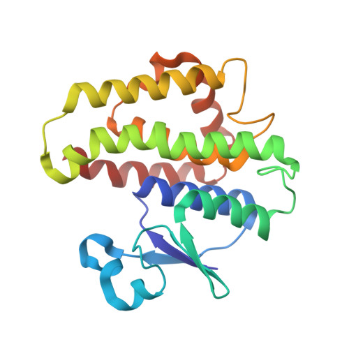
    We report four new crystal structures for Delta class glutathione transferases from insects. We compare these new structures as well as several previously reported structures to determine that structural transitions can be observed with ligand binding. These transitions occurred in the regions around the active site entrance, including alpha helix 2, C-terminus of alpha helix 4 including the loop to helix 5 and the C-terminus of helix 8. These structural movements have been reported or postulated to occur for several other glutathione transferase classes; however, this is the first report showing structural evidence of all these movements occurring, in this case in Delta class glutathione transferases. These fluctuations also can be observed occurring within a single structure as there is ligand bound in only one subunit and each subunit is undergoing different conformational transitions. The structural comparisons show reorganizations occur both pre- and post-GSH ligand binding communicated through the subunit interface of the quaternary assembly. Movements of these positions would allow 'breathing' of the active site for substrate entrance, topological rearrangement for varying substrate specificity and final product release.


  • Organizational Affiliation

    Institute of Molecular Biosciences, Mahidol University, Salaya Campus, Nakhon Pathom 73170, Thailand. wjantana@yahoo.com


Macromolecules
Find similar proteins by:  (by identity cutoff)  |  3D Structure
Entity ID: 1
MoleculeChains Sequence LengthOrganismDetailsImage
Glutathione S-transferase 1-1209Drosophila melanogasterMutation(s): 0 
Gene Names: GSTD1
EC: 2.5.1.18
UniProt
Find proteins for P20432 (Drosophila melanogaster)
Explore P20432 
Go to UniProtKB:  P20432
Entity Groups  
Sequence Clusters30% Identity50% Identity70% Identity90% Identity95% Identity100% Identity
UniProt GroupP20432
Sequence Annotations
Expand
  • Reference Sequence
Small Molecules
Ligands 1 Unique
IDChains Name / Formula / InChI Key2D Diagram3D Interactions
GSH
Query on GSH

Download Ideal Coordinates CCD File 
B [auth A]GLUTATHIONE
C10 H17 N3 O6 S
RWSXRVCMGQZWBV-WDSKDSINSA-N
Experimental Data & Validation

Experimental Data

  • Method: X-RAY DIFFRACTION
  • Resolution: 1.80 Å
  • R-Value Free: 0.200 
  • R-Value Work: 0.160 
  • R-Value Observed: 0.162 
  • Space Group: C 1 2 1
Unit Cell:
Length ( Å )Angle ( ˚ )
a = 71.352α = 90
b = 63.137β = 129.55
c = 54.24γ = 90
Software Package:
Software NamePurpose
DENZOdata reduction
SCALEPACKdata scaling
REFMACrefinement
PDB_EXTRACTdata extraction
HKL-2000data collection
HKL-2000data reduction
AMoREphasing

Structure Validation

View Full Validation Report


Ligand Structure Quality Assessment 


Entry History 

Deposition Data

Revision History  (Full details and data files)

  • Version 1.0: 2011-03-30
    Type: Initial release
  • Version 1.1: 2011-07-13
    Changes: Version format compliance
  • Version 1.2: 2015-07-08
    Changes: Database references
  • Version 1.3: 2017-11-08
    Changes: Refinement description
  • Version 1.4: 2023-11-01
    Changes: Data collection, Database references, Derived calculations, Refinement description