A Disulfide-Free Single-Domain V(L) Intrabody with Blocking Activity towards Huntingtin Reveals a Novel Mode of Epitope Recognition.
Schiefner, A., Chatwell, L., Korner, J., Neumaier, I., Colby, D.W., Volkmer, R., Wittrup, K.D., Skerra, A.(2011) J Mol Biol 414: 337-355
- PubMed: 21968397
- DOI: https://doi.org/10.1016/j.jmb.2011.09.034
- Primary Citation of Related Structures:
3LRG, 3LRH - PubMed Abstract:
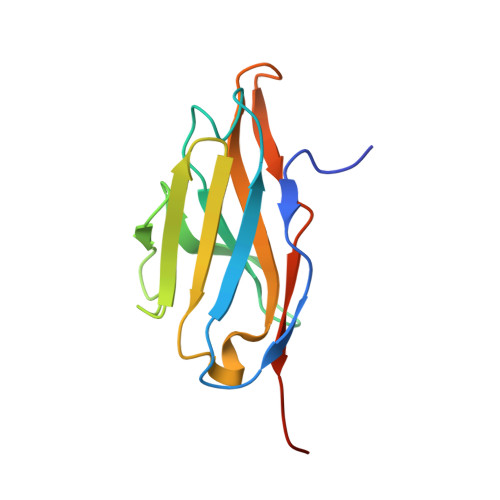
We present the crystal structure and biophysical characterization of a human V(L) [variable domain immunoglobulin (Ig) light chain] single-domain intrabody that binds to the huntingtin (Htt) protein and has been engineered for antigen recognition in the absence of its intradomain disulfide bond, otherwise conserved in the Ig fold. Analytical ultracentrifugation demonstrated that the αHtt-V(L) 12.3 domain is a stable monomer under physiological conditions even at concentrations >20 μM. Using peptide SPOT arrays, we identified the minimal binding epitope to be EKLMKAFESLKSFQ, comprising the N-terminal residues 5-18 of Htt and including the first residue of the poly-Gln stretch. X-ray structural analysis of αHtt-V(L) both as apo protein and in the presence of the epitope peptide revealed several interesting insights: first, the role of mutations acquired during the combinatorial selection process of the αHtt-V(L) 12.3 domain-initially starting from a single-chain Fv fragment-that are responsible for its stability as an individually soluble Ig domain, also lacking the disulfide bridge, and second, a previously unknown mode of antigen recognition, revealing a novel paratope. The Htt epitope peptide adopts a purely α-helical structure in the complex with αHtt-V(L) and is bound at the base of the complementarity-determining regions (CDRs) at the concave β-sheet that normally gives rise to the interface between the V(L) domain and its paired V(H) (variable domain Ig heavy chain) domain, while only few interactions with CDR-L1 and CDR-L3 are formed. Notably, this noncanonical mode of antigen binding may occur more widely in the area of in vitro selected antibody fragments, including other Ig-like scaffolds, possibly even if a V(H) domain is present.
Organizational Affiliation:
Munich Center for Integrated Protein Science and Lehrstuhl für Biologische Chemie, Technische Universität München, 85350 Freising-Weihenstephan, Germany.


