The structure of a reduced form of OxyR from Neisseria meningitidis
Sainsbury, S., Ren, J., Nettleship, J.E., Saunders, N.J., Stuart, D.I., Owens, R.J.(2010) BMC Struct Biol 10: 10-10
- PubMed: 20478059
- DOI: https://doi.org/10.1186/1472-6807-10-10
- Primary Citation of Related Structures:
3JV9 - PubMed Abstract:
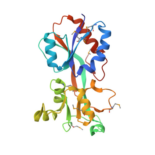
Survival of the human pathogen, Neisseria meningitidis, requires an effective response to oxidative stress resulting from the release of hydrogen peroxide by cells of the human immune system. In N. meningitidis, expression of catalase, which is responsible for detoxifying hydrogen peroxide, is controlled by OxyR, a redox responsive LysR-type regulator. OxyR responds directly to intracellular hydrogen peroxide through the reversible formation of a disulphide bond between C199 and C208 in the regulatory domain of the protein. We report the first crystal structure of the regulatory domain of an OxyR protein (NMB0173 from N. meningitidis) in the reduced state i.e. with cysteines at positions 199 and 208. The protein was crystallized under reducing conditions and the structure determined to a resolution of 2.4 A. The overall fold of the Neisseria OxyR shows a high degree of similarity to the structure of a C199S mutant OxyR from E. coli, which cannot form the redox sensitive disulphide. In the neisserial structure, C199 is located at the start of helix alpha3, separated by 18 A from C208, which is positioned between helices alpha3 and alpha4. In common with other LysR-type regulators, full length OxyR proteins are known to assemble into tetramers. Modelling of the full length neisserial OxyR as a tetramer indicated that C199 and C208 are located close to the dimer-dimer interface in the assembled tetramer. The formation of the C199-C208 disulphide may thus affect the quaternary structure of the protein. Given the high level of structural similarity between OxyR from N. meningitidis and E. coli, we conclude that the redox response mechanism is likely to be similar in both species, involving the reversible formation of a disulphide between C199-C208. Modelling suggests that disulphide formation would directly affect the interface between regulatory domains in an OxyR tetramer which in turn may lead to an alteration in the spacing/orientation of the DNA-binding domains and hence the interaction of OxyR with its DNA binding sites.
Organizational Affiliation:
The Oxford Protein Production Facility and Division of Structural Biology, Henry Wellcome Building for Genomic Medicine, University of Oxford, Roosevelt Drive, Oxford, OX3 7BN, UK.