


Crystal structure of a putative Ribokinase (Apo Form) from E.coli at 1.8A resolution.
Satyanarayana, L., Eswaramoorthy, S., Burley, S.K., Swaminathan, S.To be published.
Experimental Data Snapshot
wwPDB Validation 3D Report Full Report

Entity ID: 1 | |||||
|---|---|---|---|---|---|
| Molecule | Chains | Sequence Length | Organism | Details | Image |
| Ribokinase | 325 | Escherichia coli K-12 | Mutation(s): 0 Gene Names: b1772, JW5289, ydjH EC: 2.7.1 | 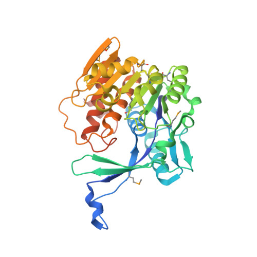 | |
UniProt | |||||
Find proteins for P77493 (Escherichia coli (strain K12)) Explore P77493 Go to UniProtKB: P77493 | |||||
Entity Groups | |||||
| Sequence Clusters | 30% Identity50% Identity70% Identity90% Identity95% Identity100% Identity | ||||
| UniProt Group | P77493 | ||||
Sequence AnnotationsExpand | |||||
| |||||
| Modified Residues 1 Unique | |||||
|---|---|---|---|---|---|
| ID | Chains | Type | Formula | 2D Diagram | Parent |
| MSE Query on MSE | A, B | L-PEPTIDE LINKING | C5 H11 N O2 Se |  | MET |
| Length ( Å ) | Angle ( ˚ ) |
|---|---|
| a = 48.088 | α = 90 |
| b = 73.718 | β = 96.49 |
| c = 82.153 | γ = 90 |
| Software Name | Purpose |
|---|---|
| CBASS | data collection |
| SHELXCD | phasing |
| SHARP | phasing |
| CNS | refinement |
| DENZO | data reduction |
| HKL-2000 | data scaling |
RCSB PDB (citation) is hosted by
RCSB PDB is a member of the

