3FSB

Crystal structure of QdtC, the dTDP-3-amino-3,6-dideoxy-D-glucose N-acetyl transferase from Thermoanaerobacterium thermosaccharolyticum in complex with CoA and dTDP-3-amino-quinovose


Experimental Data Snapshot

  • Method: X-RAY DIFFRACTION
  • Resolution: 1.95 Å
  • R-Value Free: 0.247 
  • R-Value Work: 0.183 
  • R-Value Observed: 0.185 

wwPDB Validation   3D Report Full Report


Ligand Structure Quality Assessment 


This is version 1.2 of the entry. See complete history


Literature

Structural and Functional Studies of QdtC: an N-Acetyltransferase Required for the Biosynthesis of dTDP-3-Acetamido-3,6-Dideoxy-alpha-D-Glucose.

Thoden, J.Cook, P.Schaffer, C.Messner, P.Holden, H.

(2009) Biochemistry 48: 2699-2709

  • DOI: https://doi.org/10.1021/bi802313n
  • Primary Citation of Related Structures:  
    3FS8, 3FSB, 3FSC

  • PubMed Abstract: 

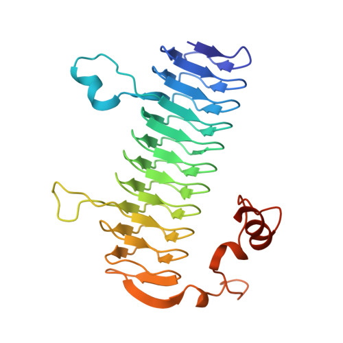
    3-Acetamido-3,6-dideoxy-alpha-D-glucose or Quip3NAc is an unusual dideoxy sugar found in the O-antigens of various Gram-negative bacteria and in the S-layer glycoprotein glycans of some Gram-positive bacteria. It is produced in these organisms as a dTDP-linked sugar, with five enzymes ultimately required for its biosynthesis. The focus of this investigation is on the enzyme QdtC, a CoA-dependent N-acetyltransferase that catalyzes the last step in the Quip3NAc biosynthetic pathway. For this analysis, three crystal structures were determined: the wild-type enzyme in the presence of acetyl-CoA and two ternary complexes of the enzyme with CoA and either dTDP-D-Quip3N or dTDP-3-amino-3,6-didexoy-alpha-D-galactose (dTDP-D-Fucp3N). Each subunit of the trimeric enzyme is dominated by a left-handed beta-helix motif with 11 turns. The three active sites are located at the subunit-subunit interfaces, and the two dTDP-sugar ligands employed in this study bind to the protein in nearly identical manners. Those residues responsible for anchoring the hexose moieties of the dTDP-sugars to the protein include Glu 141, Asn 159, and Asp 160 from one subunit and His 134 from another subunit. To probe the roles of various amino acid residues in the catalytic mechanism of the enzyme, 10 site-directed mutant proteins were constructed and their kinetic parameters measured. On the basis of these data, a catalytic mechanism is proposed for QdtC in which the acetylation of the sugar amino group does not require a catalytic base provided by the protein. Rather, the sulfur of CoA functions as the ultimate proton acceptor.


  • Organizational Affiliation

    Department of Biochemistry, University of Wisconsin, Madison, Wisconsin 53706, USA.


Macromolecules
Find similar proteins by:  (by identity cutoff)  |  3D Structure
Entity ID: 1
MoleculeChains Sequence LengthOrganismDetailsImage
QdtC
A, B
273Thermoanaerobacterium thermosaccharolyticumMutation(s): 0 
Gene Names: qdtC
UniProt
Find proteins for Q6TFC6 (Thermoanaerobacterium thermosaccharolyticum)
Explore Q6TFC6 
Go to UniProtKB:  Q6TFC6
Entity Groups  
Sequence Clusters30% Identity50% Identity70% Identity90% Identity95% Identity100% Identity
UniProt GroupQ6TFC6
Sequence Annotations
Expand
  • Reference Sequence
Small Molecules
Ligands 2 Unique
IDChains Name / Formula / InChI Key2D Diagram3D Interactions
COA
Query on COA

Download Ideal Coordinates CCD File 
C [auth A],
E [auth B]
COENZYME A
C21 H36 N7 O16 P3 S
RGJOEKWQDUBAIZ-IBOSZNHHSA-N
T3Q
Query on T3Q

Download Ideal Coordinates CCD File 
D [auth A],
F [auth B]
[(3R,4S,5S,6R)-4-amino-3,5-dihydroxy-6-methyloxan-2-yl][hydroxy-[[(2R,3S,5R)-3-hydroxy-5-(5-methyl-2,4-dioxopyrimidin-1-yl)oxolan-2-yl]methoxy]phosphoryl] hydrogen phosphate
C16 H27 N3 O14 P2
KVYJLJOGNUNRJK-HALQBZCBSA-N
Experimental Data & Validation

Experimental Data

  • Method: X-RAY DIFFRACTION
  • Resolution: 1.95 Å
  • R-Value Free: 0.247 
  • R-Value Work: 0.183 
  • R-Value Observed: 0.185 
  • Space Group: P 3
Unit Cell:
Length ( Å )Angle ( ˚ )
a = 67.6α = 90
b = 67.6β = 90
c = 112.5γ = 120
Software Package:
Software NamePurpose
PROTEUM PLUSdata collection
PHASERphasing
TNTrefinement
SAINTdata reduction
SADABSdata scaling

Structure Validation

View Full Validation Report


Ligand Structure Quality Assessment 


Entry History 

Deposition Data

Revision History  (Full details and data files)

  • Version 1.0: 2009-02-17
    Type: Initial release
  • Version 1.1: 2011-07-13
    Changes: Version format compliance
  • Version 1.2: 2023-09-06
    Changes: Data collection, Database references, Derived calculations, Refinement description