3DUV

Crystal structure of 3-deoxy-manno-octulosonate cytidylyltransferase from Haemophilus influenzae complexed with the substrate 3-deoxy-manno-octulosonate in the-configuration


Experimental Data Snapshot

  • Method: X-RAY DIFFRACTION
  • Resolution: 2.30 Å
  • R-Value Free: 0.236 
  • R-Value Work: 0.170 
  • R-Value Observed: 0.177 

wwPDB Validation   3D Report Full Report


Ligand Structure Quality Assessment 


This is version 1.2 of the entry. See complete history


Literature

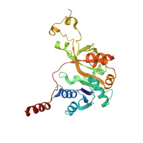
Structure of 3-deoxy-manno-octulosonate cytidylyltransferase from Haemophilus influenzae complexed with the substrate 3-deoxy-manno-octulosonate in the beta-configuration.

Yoon, H.J.Ku, M.J.Mikami, B.Suh, S.W.

(2008) Acta Crystallogr D Biol Crystallogr 64: 1292-1294

  • DOI: https://doi.org/10.1107/S0907444908036342
  • Primary Citation of Related Structures:  
    3DUV

  • PubMed Abstract: 

    The enzyme 3-deoxy-manno-octulosonate cytidylyltransferase (CMP-KDO synthetase; CKS) catalyzes the activation of 3-deoxy-D-manno-octulosonate (or 2-keto-3-deoxy-manno-octonic acid; KDO) by forming CMP-KDO. CKS is unique to Gram-negative bacteria and is an attractive target for the development of antibacterial agents. The crystal structure of CKS from Haemophilus influenzae in complex with the substrate KDO has been determined at 2.30 A resolution by combining single-wavelength anomalous diffraction and molecular-replacement methods. The two monomers in the asymmetric unit differ in the conformation of their C-terminal alpha-helix (Ala230-Asn254). The KDO bound to the active site exists as the beta-pyranose form in the (5)C(2) chair conformation. The structure of CKS from H. influenzae in complex with KDO will be useful in structure-based inhibitor design.


  • Organizational Affiliation

    Department of Chemistry, College of Natural Sciences, Seoul National University, Seoul 151-747, Republic of Korea.


Macromolecules
Find similar proteins by:  (by identity cutoff)  |  3D Structure
Entity ID: 1
MoleculeChains Sequence LengthOrganismDetailsImage
3-deoxy-manno-octulosonate cytidylyltransferase
A, B
262Haemophilus influenzaeMutation(s): 0 
Gene Names: kdsB
EC: 2.7.7.38
UniProt
Find proteins for P44490 (Haemophilus influenzae (strain ATCC 51907 / DSM 11121 / KW20 / Rd))
Explore P44490 
Go to UniProtKB:  P44490
Entity Groups  
Sequence Clusters30% Identity50% Identity70% Identity90% Identity95% Identity100% Identity
UniProt GroupP44490
Sequence Annotations
Expand
  • Reference Sequence
Small Molecules
Modified Residues  1 Unique
IDChains TypeFormula2D DiagramParent
MSE
Query on MSE
A, B
L-PEPTIDE LINKINGC5 H11 N O2 SeMET
Experimental Data & Validation

Experimental Data

  • Method: X-RAY DIFFRACTION
  • Resolution: 2.30 Å
  • R-Value Free: 0.236 
  • R-Value Work: 0.170 
  • R-Value Observed: 0.177 
  • Space Group: P 21 21 21
Unit Cell:
Length ( Å )Angle ( ˚ )
a = 48.418α = 90
b = 82.609β = 90
c = 115.712γ = 90
Software Package:
Software NamePurpose
REFMACrefinement
PDB_EXTRACTdata extraction
HKL-2000data collection
HKL-2000data reduction
HKL-2000data scaling
CNSphasing

Structure Validation

View Full Validation Report


Ligand Structure Quality Assessment 


Entry History 

Deposition Data

Revision History  (Full details and data files)

  • Version 1.0: 2008-12-09
    Type: Initial release
  • Version 1.1: 2011-07-13
    Changes: Version format compliance
  • Version 1.2: 2020-07-29
    Type: Remediation
    Reason: Carbohydrate remediation
    Changes: Advisory, Data collection, Database references, Derived calculations, Structure summary