3C8G

Crystal structure of a possible transciptional regulator YggD from Shigella flexneri 2a str. 2457T


Experimental Data Snapshot

  • Method: X-RAY DIFFRACTION
  • Resolution: 2.50 Å
  • R-Value Free: 0.270 
  • R-Value Work: 0.207 
  • R-Value Observed: 0.212 

wwPDB Validation   3D Report Full Report


This is version 1.2 of the entry. See complete history


Literature

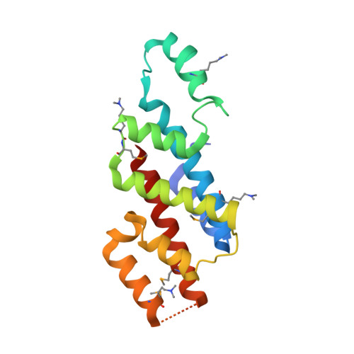
The mannitol operon repressor MtlR belongs to a new class of transcription regulators in bacteria.

Tan, K.Clancy, S.Borovilos, M.Zhou, M.Horer, S.Moy, S.Volkart, L.L.Sassoon, J.Baumann, U.Joachimiak, A.

(2009) J Biol Chem 284: 36670-36679

  • DOI: https://doi.org/10.1074/jbc.M109.062679
  • Primary Citation of Related Structures:  
    3BRJ, 3C8G

  • PubMed Abstract: 

    Many bacteria express phosphoenolpyruvate-dependent phosphotransferase systems (PTS). The mannitol-specific PTS catalyze the uptake and phosphorylation of d-mannitol. The uptake system comprises several genes encoded in the single operon. The expression of the mannitol operon is regulated by a proposed transcriptional factor, mannitol operon repressor (MtlR) that was first studied in Escherichia coli. Here we report the first crystal structures of MtlR from Vibrio parahemeolyticus (Vp-MtlR) and its homolog YggD protein from Shigella flexneri (Sf-YggD). MtlR and YggD belong to the same protein family (Pfam05068). Although Vp-MtlR and Sf-YggD share low sequence identity (22%), their overall structures are very similar, representing a novel all alpha-helical fold, and indicate similar function. However, their lack of any known DNA-binding structural motifs and their unfavorable electrostatic properties imply that MtlR/YggD are unlikely to bind a specific DNA operator directly as proposed earlier. This structural observation is further corroborated by in vitro DNA-binding studies of E. coli MtlR (Ec-MtlR), which detected no interaction of Ec-MtlR with the well characterized mannitol operator/promoter region. Therefore, MtlR/YggD belongs to a new class of transcription factors in bacteria that may regulate gene expression indirectly as a part of a larger transcriptional complex.


  • Organizational Affiliation

    Midwest Center for Structural Genomics and Structural Biology Center, Biosciences Division, Argonne National Laboratory, Argonne, Illinois 60439.


Macromolecules
Find similar proteins by:  (by identity cutoff)  |  3D Structure
Entity ID: 1
MoleculeChains Sequence LengthOrganismDetailsImage
Putative transcriptional regulator
A, C
172Shigella flexneri 2a str. 2457TMutation(s): 0 
Gene Names: yggDS3120SF2919
UniProt
Find proteins for Q83Q96 (Shigella flexneri)
Explore Q83Q96 
Go to UniProtKB:  Q83Q96
Entity Groups  
Sequence Clusters30% Identity50% Identity70% Identity90% Identity95% Identity100% Identity
UniProt GroupQ83Q96
Sequence Annotations
Expand
  • Reference Sequence
Find similar proteins by:  (by identity cutoff)  |  3D Structure
Entity ID: 2
MoleculeChains Sequence LengthOrganismDetailsImage
Putative transcriptional regulator172Shigella flexneri 2a str. 2457TMutation(s): 0 
Gene Names: yggDS3120SF2919
UniProt
Find proteins for Q83Q96 (Shigella flexneri)
Explore Q83Q96 
Go to UniProtKB:  Q83Q96
Entity Groups  
Sequence Clusters30% Identity50% Identity70% Identity90% Identity95% Identity100% Identity
UniProt GroupQ83Q96
Sequence Annotations
Expand
  • Reference Sequence
Find similar proteins by:  (by identity cutoff)  |  3D Structure
Entity ID: 3
MoleculeChains Sequence LengthOrganismDetailsImage
Putative transcriptional regulator172Shigella flexneri 2a str. 2457TMutation(s): 0 
Gene Names: yggDS3120SF2919
UniProt
Find proteins for Q83Q96 (Shigella flexneri)
Explore Q83Q96 
Go to UniProtKB:  Q83Q96
Entity Groups  
Sequence Clusters30% Identity50% Identity70% Identity90% Identity95% Identity100% Identity
UniProt GroupQ83Q96
Sequence Annotations
Expand
  • Reference Sequence
Small Molecules
Ligands 1 Unique
IDChains Name / Formula / InChI Key2D Diagram3D Interactions
ACT
Query on ACT

Download Ideal Coordinates CCD File 
E [auth A]ACETATE ION
C2 H3 O2
QTBSBXVTEAMEQO-UHFFFAOYSA-M
Modified Residues  2 Unique
IDChains TypeFormula2D DiagramParent
MLY
Query on MLY
A, C
L-PEPTIDE LINKINGC8 H18 N2 O2LYS
MSE
Query on MSE
A, C
L-PEPTIDE LINKINGC5 H11 N O2 SeMET
Experimental Data & Validation

Experimental Data

  • Method: X-RAY DIFFRACTION
  • Resolution: 2.50 Å
  • R-Value Free: 0.270 
  • R-Value Work: 0.207 
  • R-Value Observed: 0.212 
  • Space Group: C 1 2 1
Unit Cell:
Length ( Å )Angle ( ˚ )
a = 71.365α = 90
b = 70.946β = 90.43
c = 159.037γ = 90
Software Package:
Software NamePurpose
REFMACrefinement
SBC-Collectdata collection
HKL-3000data reduction
HKL-3000data scaling
SHELXDphasing
MLPHAREphasing
DMphasing
RESOLVEphasing
HKL-3000phasing

Structure Validation

View Full Validation Report


Entry History 

Deposition Data

Revision History  (Full details and data files)

  • Version 1.0: 2008-02-19
    Type: Initial release
  • Version 1.1: 2011-07-13
    Changes: Advisory, Version format compliance
  • Version 1.2: 2017-11-01
    Changes: Derived calculations