3BAZ

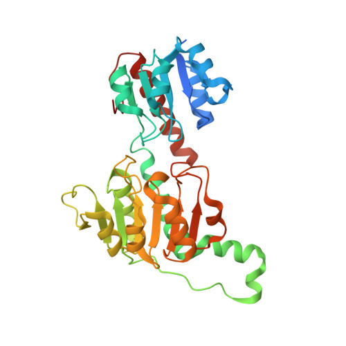
Structure of hydroxyphenylpyruvate reductase from coleus blumei in complex with NADP+


Experimental Data Snapshot

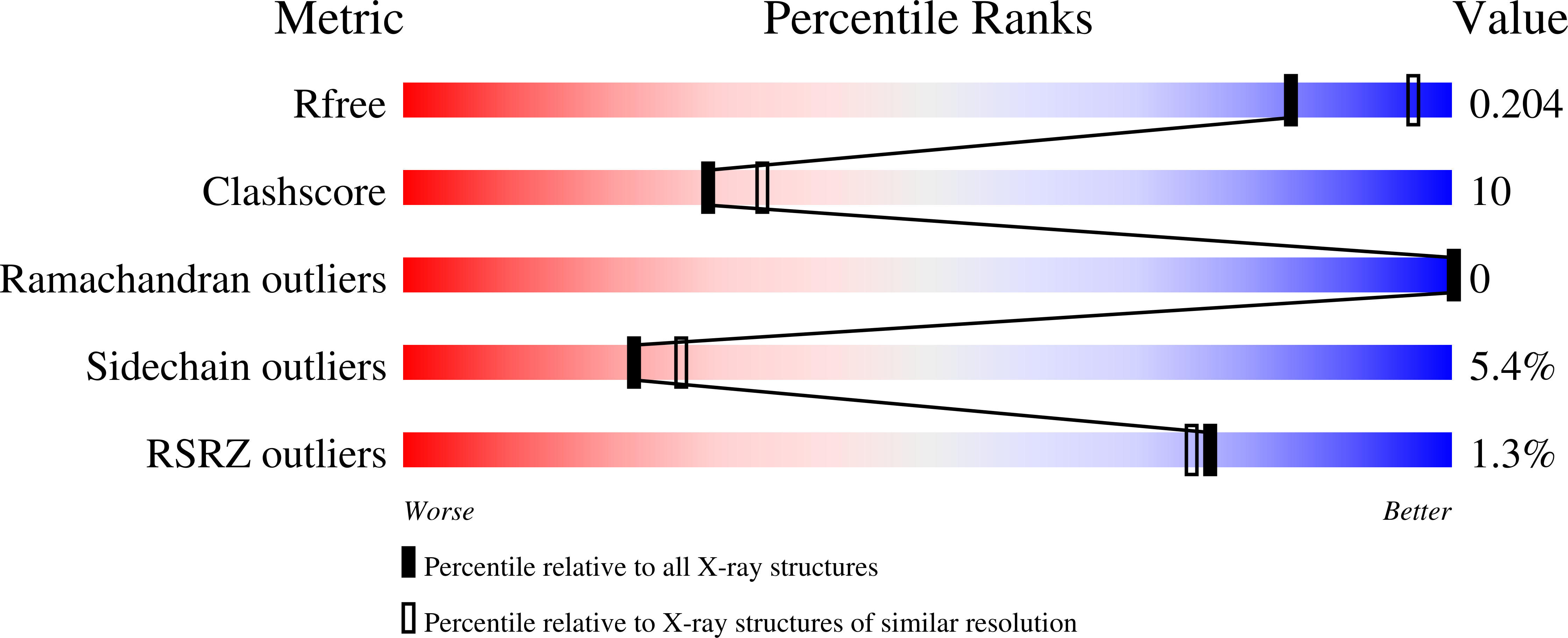
  • Method: X-RAY DIFFRACTION
  • Resolution: 2.20 Å
  • R-Value Free: 0.260 
  • R-Value Work: 0.211 
  • R-Value Observed: 0.211 

wwPDB Validation   3D Report Full Report


Ligand Structure Quality Assessment 


This is version 1.3 of the entry. See complete history


Literature

Structure and substrate docking of a hydroxy(phenyl)pyruvate reductase from the higher plant Coleus blumei Benth

Janiak, V.Petersen, M.Zentgraf, M.Klebe, G.Heine, A.

(2010) Acta Crystallogr D Biol Crystallogr 66: 593-603

  • DOI: https://doi.org/10.1107/S0907444910006360
  • Primary Citation of Related Structures:  
    3BA1, 3BAZ

  • PubMed Abstract: 

    Hydroxy(phenyl)pyruvate reductase [H(P)PR] belongs to the family of D-isomer-specific 2-hydroxyacid dehydrogenases and catalyzes the reduction of hydroxyphenylpyruvates as well as hydroxypyruvate and pyruvate to the corresponding lactates. Other non-aromatic substrates are also accepted. NADPH is the preferred cosubstrate. The crystal structure of the enzyme from Coleus blumei (Lamiaceae) has been determined at 1.47 A resolution. In addition to the apoenzyme, the structure of a complex with NADP(+) was determined at a resolution of 2.2 A. H(P)PR is a dimer with a molecular mass of 34 113 Da per subunit. The structure is similar to those of other members of the enzyme family and consists of two domains separated by a deep catalytic cleft. To gain insights into substrate binding, several compounds were docked into the cosubstrate complex structure using the program AutoDock. The results show two possible binding modes with similar docking energy. However, only binding mode A provides the necessary environment in the active centre for hydride and proton transfer during reduction, leading to the formation of the (R)-enantiomer of lactate and/or hydroxyphenyllactate.


  • Organizational Affiliation

    Institut für Pharmazeutische Biologie, Philipps-Universität Marburg, Deutschhausstrasse 17A, 35037 Marburg, Germany.


Macromolecules
Find similar proteins by:  (by identity cutoff)  |  3D Structure
Entity ID: 1
MoleculeChains Sequence LengthOrganismDetailsImage
Hydroxyphenylpyruvate reductase333Coleus scutellarioidesMutation(s): 0 
Gene Names: hppr
EC: 1.1.1.237
UniProt
Find proteins for Q65CJ7 (Plectranthus scutellarioides)
Explore Q65CJ7 
Go to UniProtKB:  Q65CJ7
Entity Groups  
Sequence Clusters30% Identity50% Identity70% Identity90% Identity95% Identity100% Identity
UniProt GroupQ65CJ7
Sequence Annotations
Expand
  • Reference Sequence
Small Molecules
Ligands 1 Unique
IDChains Name / Formula / InChI Key2D Diagram3D Interactions
NAP
Query on NAP

Download Ideal Coordinates CCD File 
B [auth A]NADP NICOTINAMIDE-ADENINE-DINUCLEOTIDE PHOSPHATE
C21 H28 N7 O17 P3
XJLXINKUBYWONI-NNYOXOHSSA-N
Experimental Data & Validation

Experimental Data

  • Method: X-RAY DIFFRACTION
  • Resolution: 2.20 Å
  • R-Value Free: 0.260 
  • R-Value Work: 0.211 
  • R-Value Observed: 0.211 
  • Space Group: P 41 21 2
Unit Cell:
Length ( Å )Angle ( ˚ )
a = 63.5α = 90
b = 63.5β = 90
c = 222.6γ = 90
Software Package:
Software NamePurpose
SHELXmodel building
SHELXL-97refinement
CrystalCleardata collection
HKL-2000data reduction
HKL-2000data scaling
PHASERphasing

Structure Validation

View Full Validation Report


Ligand Structure Quality Assessment 


Entry History 

Deposition Data

Revision History  (Full details and data files)

  • Version 1.0: 2008-11-11
    Type: Initial release
  • Version 1.1: 2011-07-13
    Changes: Version format compliance
  • Version 1.2: 2012-04-25
    Changes: Database references
  • Version 1.3: 2023-11-01
    Changes: Data collection, Database references, Derived calculations, Refinement description