3B5B

Crystal structure of the thymidylate synthase k48q


Experimental Data Snapshot

  • Method: X-RAY DIFFRACTION
  • Resolution: 2.70 Å
  • R-Value Free: 0.234 
  • R-Value Work: 0.193 

wwPDB Validation   3D Report Full Report


This is version 1.6 of the entry. See complete history


Literature

Role of an invariant lysine residue in folate binding on Escherichia coli thymidylate synthase: Calorimetric and crystallographic analysis of the K48Q mutant.

Arvizu-Flores, A.A.Sugich-Miranda, R.Arreola, R.Garcia-Orozco, K.D.Velazquez-Contreras, E.F.Montfort, W.R.Maley, F.Sotelo-Mundo, R.R.

(2008) Int J Biochem Cell Biol 40: 2206-2217

  • DOI: https://doi.org/10.1016/j.biocel.2008.02.025
  • Primary Citation of Related Structures:  
    2VET, 2VF0, 3B5B

  • PubMed Abstract: 

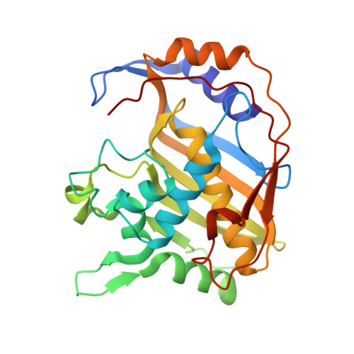
    Thymidylate synthase (TS) catalyzes the reductive methylation of deoxyuridine monophosphate (dUMP) using methylene tetrahydrofolate (CH(2)THF) as cofactor, the glutamate tail of which forms a water-mediated hydrogen bond with an invariant lysine residue of this enzyme. To understand the role of this interaction, we studied the K48Q mutant of Escherichia coli TS using structural and biophysical methods. The k(cat) of the K48Q mutant was 430-fold lower than wild-type TS in activity, while the K(m) for the (R)-stereoisomer of CH(2)THF was 300 microM, about 30-fold larger than K(m) from the wild-type TS. Affinity constants were determined using isothermal titration calorimetry, which showed that binding was reduced by one order of magnitude for folate-like TS inhibitors, such as propargyl-dideazafolate (PDDF) or compounds that distort the TS active site like BW1843U89 (U89). The crystal structure of the K48Q-dUMP complex revealed that dUMP binding is not impaired in the mutant, and that U89 in a ternary complex of K48Q-nucleotide-U89 was bound in the active site with subtle differences relative to comparable wild-type complexes. PDDF failed to form ternary complexes with K48Q and dUMP. Thermodynamic data correlated with the structural determinations, since PDDF binding was dominated by enthalpic effects while U89 had an important entropic component. In conclusion, K48 is critical for catalysis since it leads to a productive CH(2)THF binding, while mutation at this residue does not affect much the binding of inhibitors that do not make contact with this group.


  • Organizational Affiliation

    Aquatic Molecular Biology Laboratory, Centro de Investigación en Alimentación y Desarrollo, A.C. Hermosillo, Sonora 83000, Mexico.


Macromolecules
Find similar proteins by:  (by identity cutoff)  |  3D Structure
Entity ID: 1
MoleculeChains Sequence LengthOrganismDetailsImage
Thymidylate synthase
A, B
264Escherichia coli BL21Mutation(s): 1 
Gene Names: thyA
EC: 2.1.1.45
UniProt
Find proteins for P0A884 (Escherichia coli (strain K12))
Explore P0A884 
Go to UniProtKB:  P0A884
Entity Groups  
Sequence Clusters30% Identity50% Identity70% Identity90% Identity95% Identity100% Identity
UniProt GroupP0A884
Sequence Annotations
Expand
  • Reference Sequence
Small Molecules
Ligands 3 Unique
IDChains Name / Formula / InChI Key2D Diagram3D Interactions
NDU
Query on NDU

Download Ideal Coordinates CCD File 
C [auth A]2'-DEOXY-5-NITROURIDINE 5'-MONOPHOSPHATE
C9 H14 N3 O10 P
ZYBJIJYGXJSDTC-XZBKPIIZSA-N
NDN
Query on NDN

Download Ideal Coordinates CCD File 
E [auth B]2'-DEOXY-5-NITROURIDINE 5'-(DIHYDROGEN PHOSPHATE)
C9 H12 N3 O10 P
LVLPVLNNXRTCHP-RRKCRQDMSA-N
FMT
Query on FMT

Download Ideal Coordinates CCD File 
D [auth A],
F [auth B]
FORMIC ACID
C H2 O2
BDAGIHXWWSANSR-UHFFFAOYSA-N
Experimental Data & Validation

Experimental Data

  • Method: X-RAY DIFFRACTION
  • Resolution: 2.70 Å
  • R-Value Free: 0.234 
  • R-Value Work: 0.193 
  • Space Group: P 63
Unit Cell:
Length ( Å )Angle ( ˚ )
a = 127.37α = 90
b = 127.37β = 90
c = 67.84γ = 120
Software Package:
Software NamePurpose
CNSrefinement
REFMACrefinement
PDB_EXTRACTdata extraction
MADNESSdata collection
PROCORdata reduction
Agrovatadata scaling
ROTAVATAdata scaling
TRUNCATEdata scaling
X-PLORphasing
SCALAdata scaling

Structure Validation

View Full Validation Report


Entry History 

Deposition Data

Revision History  (Full details and data files)

  • Version 1.0: 2007-12-25
    Type: Initial release
  • Version 1.1: 2011-07-13
    Changes: Non-polymer description, Version format compliance
  • Version 1.2: 2017-10-25
    Changes: Refinement description
  • Version 1.3: 2018-04-18
    Changes: Data collection
  • Version 1.4: 2019-07-24
    Changes: Data collection, Derived calculations, Refinement description
  • Version 1.5: 2021-10-20
    Changes: Database references, Derived calculations
  • Version 1.6: 2023-08-30
    Changes: Data collection, Refinement description