Crystal structures of the lumazine protein from Photobacterium kishitanii in complexes with the authentic chromophore, 6,7-dimethyl-8-(1'-D-ribityl) lumazine and its analogues, riboflavin and FMN, at high resolution
Sato, Y., Shimizu, S., Ohtaki, A., Noguchi, K., Miyatake, H., Dohmae, N., Sasaki, S., Odaka, M., Yohda, M.(2009) J Bacteriol 192: 127-133
- PubMed: 19854891
- DOI: https://doi.org/10.1128/JB.01015-09
- Primary Citation of Related Structures:
3A35, 3A3B, 3A3G - PubMed Abstract:
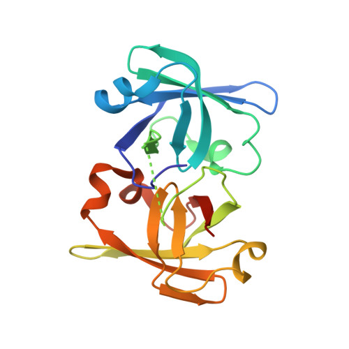
Lumazine protein (LumP) is a fluorescent accessory protein having 6,7-dimethyl-8-(1'-d-ribityl) lumazine (DMRL) as its authentic chromophore. It modulates the emission of bacterial luciferase to shorter wavelengths with increasing luminous strength. To obtain structural information on the native structure as well as the interaction with bacterial luciferase, we have determined the crystal structures of LumP from Photobacterium kishitanii in complexes with DMRL and its analogues, riboflavin (RBF) and flavin mononucleotide (FMN), at resolutions of 2.00, 1.42, and 2.00 A. LumP consists of two beta barrels that have nearly identical folds, the N-terminal and C-terminal barrels. The structures of LumP in complex with all of the chromophores studied are all essentially identical, except around the chromophores. In all of the structures, the chromophore is tethered to the narrow cavity via many hydrogen bonds in the N-terminal domain. These are absent in the C-terminal domain. Hydrogen bonding in LumP-FMN is decreased in comparison with that in LumP-RBF because the phosphate moiety of FMN protrudes out of the narrow cavity. In LumP-DMRL, the side chain of Gln65 is close to the ring system, and a new water molecule that stabilizes the ligand is observed near Ser48. Therefore, DMRL packs more tightly in the ligand-binding site than RBF or FMN. A docking simulation of bacterial luciferase and LumP suggests that the chromophore is located close enough for direct energy transfer to occur. Moreover, the surface potentials around the ligand-binding sites of LumP and bacterial luciferase exhibit complementary charge distributions, which would have a significant effect on the interaction between LumP and luciferase.
Organizational Affiliation:
Department of Biotechnology and Life Science, Tokyo University of Agriculture and Technology, 2-24-16 Naka-cho, Koganei, Tokyo 184-8588, Japan.