2Z7G

Crystal structure of adenosine deaminase ligated with EHNA


Experimental Data Snapshot

  • Method: X-RAY DIFFRACTION
  • Resolution: 2.52 Å
  • R-Value Free: 0.247 
  • R-Value Work: 0.207 
  • R-Value Observed: 0.210 

wwPDB Validation   3D Report Full Report


Ligand Structure Quality Assessment 


This is version 1.2 of the entry. See complete history


Literature

Conformational change of adenosine deaminase during ligand-exchange in a crystal

Kinoshita, T.Tada, T.Nakanishi, I.

(2008) Biochem Biophys Res Commun 373: 53-57

  • DOI: https://doi.org/10.1016/j.bbrc.2008.05.180
  • Primary Citation of Related Structures:  
    2Z7G

  • PubMed Abstract: 

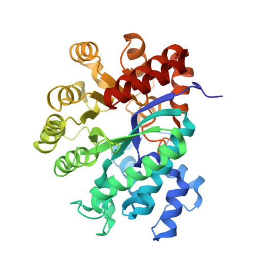
    Adenosine deaminase (ADA) perpetuates chronic inflammation by degrading extracellular adenosine which is toxic for lymphocytes. ADA has two distinct conformations: open form and closed form. From the crystal structures with various ligands, the non-nucleoside type inhibitors bind to the active site occupying the critical water-binding-position and sustain the open form of apo-ADA. In contrast, substrate mimics do not occupy the critical position, and induce the large conformational change to the closed form. However, it is difficult to predict the binding of (+)-erythro-9-(2-hydroxy-3-nonyl)adenine (EHNA), as it possesses characteristic parts of both the substrate and the non-nucleoside inhibitors. The crystal structure shows that EHNA binds to the open form through a novel recognition of the adenine base accompanying conformational change from the closed form of the PR-ADA complex in crystalline state.


  • Organizational Affiliation

    Graduate School of Science, Osaka Prefecture University, Gakuencho 1-1, Sakai, Osaka 599-8531, Japan. kinotk@b.s.osakafu-u.ac.jp


Macromolecules
Find similar proteins by:  (by identity cutoff)  |  3D Structure
Entity ID: 1
MoleculeChains Sequence LengthOrganismDetailsImage
Adenosine deaminase356Bos taurusMutation(s): 0 
EC: 3.5.4.4
UniProt
Find proteins for P56658 (Bos taurus)
Explore P56658 
Go to UniProtKB:  P56658
Entity Groups  
Sequence Clusters30% Identity50% Identity70% Identity90% Identity95% Identity100% Identity
UniProt GroupP56658
Sequence Annotations
Expand
  • Reference Sequence
Small Molecules
Ligands 2 Unique
IDChains Name / Formula / InChI Key2D Diagram3D Interactions
EH9
Query on EH9

Download Ideal Coordinates CCD File 
C [auth A](2S,3R)-3-(6-amino-9H-purin-9-yl)nonan-2-ol
C14 H23 N5 O
IOSAAWHGJUZBOG-WDEREUQCSA-N
ZN
Query on ZN

Download Ideal Coordinates CCD File 
B [auth A]ZINC ION
Zn
PTFCDOFLOPIGGS-UHFFFAOYSA-N
Binding Affinity Annotations 
IDSourceBinding Affinity
EH9 BindingDB:  2Z7G Ki: min: 0.82, max: 1140 (nM) from 10 assay(s)
PDBBind:  2Z7G Ki: 1.13 (nM) from 1 assay(s)
Experimental Data & Validation

Experimental Data

  • Method: X-RAY DIFFRACTION
  • Resolution: 2.52 Å
  • R-Value Free: 0.247 
  • R-Value Work: 0.207 
  • R-Value Observed: 0.210 
  • Space Group: P 43 21 2
Unit Cell:
Length ( Å )Angle ( ˚ )
a = 77.1α = 90
b = 77.1β = 90
c = 135.66γ = 90
Software Package:
Software NamePurpose
CrystalCleardata collection
MOLREPphasing
CNXrefinement
CrystalCleardata reduction
CrystalCleardata scaling

Structure Validation

View Full Validation Report


Ligand Structure Quality Assessment 


Entry History 

Deposition Data

Revision History  (Full details and data files)

  • Version 1.0: 2008-07-01
    Type: Initial release
  • Version 1.1: 2011-07-13
    Changes: Version format compliance
  • Version 1.2: 2023-11-01
    Changes: Data collection, Database references, Derived calculations, Refinement description, Structure summary