2XZE

Structural basis for AMSH-ESCRT-III CHMP3 interaction


Experimental Data Snapshot

  • Method: X-RAY DIFFRACTION
  • Resolution: 1.75 Å
  • R-Value Free: 0.227 
  • R-Value Work: 0.192 
  • R-Value Observed: 0.193 

wwPDB Validation   3D Report Full Report


This is version 1.0 of the entry. See complete history


Literature

Structural Basis for Escrt-III Chmp3 Recruitment of Amsh.

Solomons, J.Sabin, C.Poudevigne, E.Usami, Y.Hulsik, D.L.Macheboeuf, P.Hartlieb, B.Gottlinger, H.Weissenhorn, W.

(2011) Structure 19: 1149

  • DOI: https://doi.org/10.1016/j.str.2011.05.011
  • Primary Citation of Related Structures:  
    2XZE

  • PubMed Abstract: 

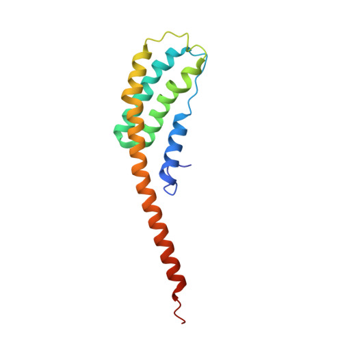
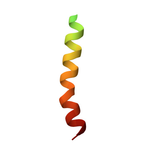
    Endosomal sorting complexes required for transport (ESCRT) recognize ubiquitinated cargo and catalyze diverse budding processes including multivesicular body biogenesis, enveloped virus egress, and cytokinesis. We present the crystal structure of an N-terminal fragment of the deubiquitinating enzyme AMSH (AMSHΔC) in complex with the C-terminal region of ESCRT-III CHMP3 (CHMP3ΔN). AMSHΔC folds into an elongated 90 Å long helical assembly that includes an unusual MIT domain. CHMP3ΔN is unstructured in solution and helical in complex with AMSHΔC, revealing a novel MIT domain interacting motif (MIM) that does not overlap with the CHMP1-AMSH binding site. ITC and SPR measurements demonstrate an unusual high-affinity MIM-MIT interaction. Structural analysis suggests a regulatory role for the N-terminal helical segment of AMSHΔC and its destabilization leads to a loss of function during HIV-1 budding. Our results indicate a tight coupling of ESCRT-III CHMP3 and AMSH functions and provide insight into the regulation of ESCRT-III.


  • Organizational Affiliation

    Unit of Virus Host Cell Interactions (UVHCI) UMI 3265 Université Joseph Fourier-EMBL-CNRS, 6 rue Jules Horowitz 38042 Grenoble Cedex 9, France.


Macromolecules
Find similar proteins by:  (by identity cutoff)  |  3D Structure
Entity ID: 1
MoleculeChains Sequence LengthOrganismDetailsImage
STAM-BINDING PROTEIN
A, B
146Homo sapiensMutation(s): 0 
EC: 3.4.19
UniProt & NIH Common Fund Data Resources
Find proteins for O95630 (Homo sapiens)
Explore O95630 
Go to UniProtKB:  O95630
PHAROS:  O95630
GTEx:  ENSG00000124356 
Entity Groups  
Sequence Clusters30% Identity50% Identity70% Identity90% Identity95% Identity100% Identity
UniProt GroupO95630
Sequence Annotations
Expand
  • Reference Sequence
Find similar proteins by:  (by identity cutoff)  |  3D Structure
Entity ID: 2
MoleculeChains Sequence LengthOrganismDetailsImage
CHARGED MULTIVESICULAR BODY PROTEIN 3C [auth Q],
D [auth R]
40Homo sapiensMutation(s): 0 
UniProt & NIH Common Fund Data Resources
Find proteins for Q9Y3E7 (Homo sapiens)
Explore Q9Y3E7 
Go to UniProtKB:  Q9Y3E7
PHAROS:  Q9Y3E7
GTEx:  ENSG00000115561 
Entity Groups  
Sequence Clusters30% Identity50% Identity70% Identity90% Identity95% Identity100% Identity
UniProt GroupQ9Y3E7
Sequence Annotations
Expand
  • Reference Sequence
Experimental Data & Validation

Experimental Data

  • Method: X-RAY DIFFRACTION
  • Resolution: 1.75 Å
  • R-Value Free: 0.227 
  • R-Value Work: 0.192 
  • R-Value Observed: 0.193 
  • Space Group: P 41
Unit Cell:
Length ( Å )Angle ( ˚ )
a = 45.97α = 90
b = 45.97β = 90
c = 206.91γ = 90
Software Package:
Software NamePurpose
REFMACrefinement
MOSFLMdata reduction
SCALAdata scaling
Auto-Rickshawphasing

Structure Validation

View Full Validation Report


Entry History 

Deposition Data

Revision History  (Full details and data files)

  • Version 1.0: 2011-08-24
    Type: Initial release