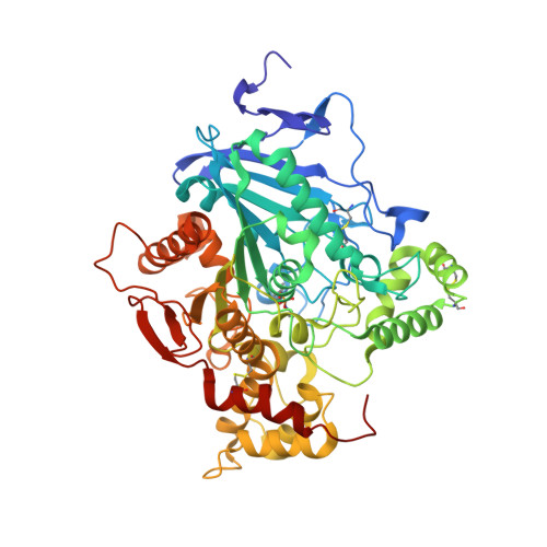
2WU3

CRYSTAL STRUCTURE OF MOUSE ACETYLCHOLINESTERASE IN COMPLEX WITH FENAMIPHOS AND HI-6


Experimental Data Snapshot

  • Method: X-RAY DIFFRACTION
  • Resolution: 2.70 Å
  • R-Value Free: 0.226 
  • R-Value Work: 0.171 
  • R-Value Observed: 0.172 

wwPDB Validation   3D Report Full Report


Ligand Structure Quality Assessment 


This is version 2.3 of the entry. See complete history


Literature

Crystal Structures of Oxime-Bound Fenamiphos-Acetylcholinesterases: Reactivation Involving Flipping of the His447 Ring to Form a Reactive Glu334-His447-Oxime Triad.

Hornberg, A.Artursson, E.Warme, R.Pang, Y.-P.Ekstrom, F.

(2010) Biochem Pharmacol 79: 507

  • DOI: https://doi.org/10.1016/j.bcp.2009.08.027
  • Primary Citation of Related Structures:  
    2WU3, 2WU4

  • PubMed Abstract: 

    Organophosphorus insecticides and nerve agents inhibit the vital enzyme acetylcholinesterase by covalently bonding to the catalytic serine residue of the enzyme. Oxime-based reactivators, such as [(E)-[1-[(4-carbamoylpyridin-1-ium-1-yl)methoxymethyl]pyridin-2-ylidene]methyl]-oxoazanium dichloride (HI-6) and 1,7-heptylene-bis-N,N'-2-pyridiniumaldoxime dichloride (Ortho-7), restore the organophosphate-inhibited enzymatic activity by cleaving the phosphorous conjugate. In this article, we report the intermolecular interactions between Mus musculus acetylcholinesterase inhibited by the insecticide fenamiphos (fep-mAChE) and HI-6 or Ortho-7 revealed by a combination of crystallography and kinetics. The crystal structures of the two oxime-bound fep-mAChE complexes show that both oximes interact with the peripheral anionic site involving different conformations of Trp286 and different peripheral-site residues (Tyr124 for HI-6 and Tyr72 for Ortho-7). Moreover, residues at catalytic site of the HI-6-bound fep-mAChE complex adopt conformations that are similar to those in the apo mAChE, whereas significant conformational changes are observed for the corresponding residues in the Ortho-7-bound fep-mAChE complex. Interestingly, flipping of the His447 imidazole ring allows the formation of a hydrogen bonding network among the Glu334-His447-Ortho-7 triad, which presumably deprotonates the Ortho-7 oxime hydroxyl group, increases the nucleophilicity of the oxime group, and leads to cleavage of the phosphorous conjugate. These results offer insights into a detailed reactivation mechanism for the oximes and development of improved reactivators.


  • Organizational Affiliation

    Swedish Defence Research Agency, Division of CBRN Defence and Security, S-901 82 Umeå, Sweden.


Macromolecules
Find similar proteins by:  (by identity cutoff)  |  3D Structure
Entity ID: 1
MoleculeChains Sequence LengthOrganismDetailsImage
ACETYLCHOLINESTERASE
A, B
548Mus musculusMutation(s): 0 
EC: 3.1.1.7
UniProt & NIH Common Fund Data Resources
Find proteins for P21836 (Mus musculus)
Explore P21836 
Go to UniProtKB:  P21836
IMPC:  MGI:87876
Entity Groups  
Sequence Clusters30% Identity50% Identity70% Identity90% Identity95% Identity100% Identity
UniProt GroupP21836
Sequence Annotations
Expand
  • Reference Sequence
Small Molecules
Ligands 4 Unique
IDChains Name / Formula / InChI Key2D Diagram3D Interactions
HI6
Query on HI6

Download Ideal Coordinates CCD File 
C [auth A],
H [auth B]
4-(AMINOCARBONYL)-1-[({2-[(E)-(HYDROXYIMINO)METHYL]PYRIDINIUM-1-YL}METHOXY)METHYL]PYRIDINIUM
C14 H16 N4 O3
FJZDLOMCEPUCII-UHFFFAOYSA-P
P6G
Query on P6G

Download Ideal Coordinates CCD File 
F [auth A]HEXAETHYLENE GLYCOL
C12 H26 O7
IIRDTKBZINWQAW-UHFFFAOYSA-N
NAG
Query on NAG

Download Ideal Coordinates CCD File 
D [auth A],
E [auth A]
2-acetamido-2-deoxy-beta-D-glucopyranose
C8 H15 N O6
OVRNDRQMDRJTHS-FMDGEEDCSA-N
CO3
Query on CO3

Download Ideal Coordinates CCD File 
G [auth A],
I [auth B]
CARBONATE ION
C O3
BVKZGUZCCUSVTD-UHFFFAOYSA-L
Modified Residues  1 Unique
IDChains TypeFormula2D DiagramParent
SXE
Query on SXE
A, B
L-PEPTIDE LINKINGC8 H19 N2 O5 PSER
Binding Affinity Annotations 
IDSourceBinding Affinity
HI6 BindingDB:  2WU3 Kd: min: 1.50e+4, max: 2.34e+5 (nM) from 6 assay(s)
IC50: min: 6.36e+5, max: 1.36e+6 (nM) from 3 assay(s)
Experimental Data & Validation

Experimental Data

  • Method: X-RAY DIFFRACTION
  • Resolution: 2.70 Å
  • R-Value Free: 0.226 
  • R-Value Work: 0.171 
  • R-Value Observed: 0.172 
  • Space Group: P 21 21 21
Unit Cell:
Length ( Å )Angle ( ˚ )
a = 78.76α = 90
b = 111.23β = 90
c = 227.7γ = 90
Software Package:
Software NamePurpose
PHENIXrefinement
XDSdata reduction
SCALAdata scaling
CCP4phasing

Structure Validation

View Full Validation Report


Ligand Structure Quality Assessment 


Entry History 

Deposition Data

Revision History  (Full details and data files)

  • Version 1.0: 2009-10-20
    Type: Initial release
  • Version 1.1: 2011-05-08
    Changes: Version format compliance
  • Version 1.2: 2011-07-13
    Changes: Version format compliance
  • Version 1.3: 2017-07-12
    Changes: Advisory, Data collection, Derived calculations
  • Version 2.0: 2019-05-15
    Changes: Data collection, Derived calculations, Polymer sequence
  • Version 2.1: 2020-07-29
    Type: Remediation
    Reason: Carbohydrate remediation
    Changes: Data collection, Derived calculations, Other, Structure summary
  • Version 2.2: 2021-05-12
    Changes: Derived calculations, Structure summary
  • Version 2.3: 2023-12-20
    Changes: Data collection, Database references, Refinement description