2VSY

Xanthomonas campestris putative OGT (XCC0866), apostructure


Experimental Data Snapshot

  • Method: X-RAY DIFFRACTION
  • Resolution: 2.10 Å
  • R-Value Free: 0.236 
  • R-Value Work: 0.193 
  • R-Value Observed: 0.194 

wwPDB Validation   3D Report Full Report


This is version 1.1 of the entry. See complete history


Literature

Structural Insights Into Mechanism and Specificity of O-Glcnac Transferase.

Clarke, A.J.Hurtado-Guerrero, R.Pathak, S.Schuttelkopf, A.W.Borodkin, V.Shepherd, S.M.Ibrahim, A.F.M.Van Aalten, D.M.F.

(2008) EMBO J 27: 2780

  • DOI: https://doi.org/10.1038/emboj.2008.186
  • Primary Citation of Related Structures:  
    2JLB, 2VSY

  • PubMed Abstract: 

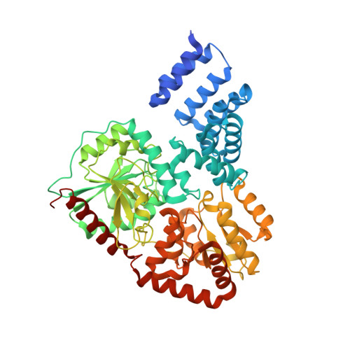
    Post-translational modification of protein serines/threonines with N-acetylglucosamine (O-GlcNAc) is dynamic, inducible and abundant, regulating many cellular processes by interfering with protein phosphorylation. O-GlcNAcylation is regulated by O-GlcNAc transferase (OGT) and O-GlcNAcase, both encoded by single, essential, genes in metazoan genomes. It is not understood how OGT recognises its sugar nucleotide donor and performs O-GlcNAc transfer onto proteins/peptides, and how the enzyme recognises specific cellular protein substrates. Here, we show, by X-ray crystallography and mutagenesis, that OGT adopts the (metal-independent) GT-B fold and binds a UDP-GlcNAc analogue at the bottom of a highly conserved putative peptide-binding groove, covered by a mobile loop. Strikingly, the tetratricopeptide repeats (TPRs) tightly interact with the active site to form a continuous 120 A putative interaction surface, whereas the previously predicted phosphatidylinositide-binding site locates to the opposite end of the catalytic domain. On the basis of the structure, we identify truncation/point mutants of the TPRs that have differential effects on activity towards proteins/peptides, giving first insights into how OGT may recognise its substrates.


  • Organizational Affiliation

    Division of Biological Chemistry & Drug Discovery, College of Life Sciences, University of Dundee, Dundee, UK.


Macromolecules
Find similar proteins by:  (by identity cutoff)  |  3D Structure
Entity ID: 1
MoleculeChains Sequence LengthOrganismDetailsImage
XCC0866
A, B
568Xanthomonas campestris pv. campestris str. ATCC 33913Mutation(s): 0 
UniProt
Find proteins for Q8PC69 (Xanthomonas campestris pv. campestris (strain ATCC 33913 / DSM 3586 / NCPPB 528 / LMG 568 / P 25))
Explore Q8PC69 
Go to UniProtKB:  Q8PC69
Entity Groups  
Sequence Clusters30% Identity50% Identity70% Identity90% Identity95% Identity100% Identity
UniProt GroupQ8PC69
Sequence Annotations
Expand
  • Reference Sequence
Small Molecules
Ligands 4 Unique
IDChains Name / Formula / InChI Key2D Diagram3D Interactions
NHE
Query on NHE

Download Ideal Coordinates CCD File 
C [auth A],
H [auth B],
I [auth B]
2-[N-CYCLOHEXYLAMINO]ETHANE SULFONIC ACID
C8 H17 N O3 S
MKWKNSIESPFAQN-UHFFFAOYSA-N
PR
Query on PR

Download Ideal Coordinates CCD File 
F [auth A],
G [auth A],
L [auth B]
PRASEODYMIUM ION
Pr
WCWKKSOQLQEJTE-UHFFFAOYSA-N
PEG
Query on PEG

Download Ideal Coordinates CCD File 
D [auth A],
E [auth A]
DI(HYDROXYETHYL)ETHER
C4 H10 O3
MTHSVFCYNBDYFN-UHFFFAOYSA-N
CL
Query on CL

Download Ideal Coordinates CCD File 
J [auth B],
K [auth B]
CHLORIDE ION
Cl
VEXZGXHMUGYJMC-UHFFFAOYSA-M
Experimental Data & Validation

Experimental Data

  • Method: X-RAY DIFFRACTION
  • Resolution: 2.10 Å
  • R-Value Free: 0.236 
  • R-Value Work: 0.193 
  • R-Value Observed: 0.194 
  • Space Group: P 21 21 21
Unit Cell:
Length ( Å )Angle ( ˚ )
a = 81.523α = 90
b = 100.096β = 90
c = 156.274γ = 90
Software Package:
Software NamePurpose
REFMACrefinement
DENZOdata reduction
SCALEPACKdata scaling
AMoREphasing

Structure Validation

View Full Validation Report


Entry History 

Deposition Data

Revision History  (Full details and data files)

  • Version 1.0: 2008-11-18
    Type: Initial release
  • Version 1.1: 2011-07-13
    Changes: Advisory, Version format compliance