2V7F

Structure of P. abyssi RPS19 protein


Experimental Data Snapshot

  • Method: X-RAY DIFFRACTION
  • Resolution: 1.15 Å
  • R-Value Free: 0.155 
  • R-Value Work: 0.137 
  • R-Value Observed: 0.138 

wwPDB Validation   3D Report Full Report


This is version 1.2 of the entry. See complete history


Literature

Molecular Basis of Diamond Blackfan Anemia: Structure and Function Analysis of Rps19.

Gregory, L.A.Aguissa-Toure, A.H.Pinaud, N.Legrand, P.Gleizes, P.E.Fribourg, S.

(2007) Nucleic Acids Res 35: 5913

  • DOI: https://doi.org/10.1093/nar/gkm626
  • Primary Citation of Related Structures:  
    2V7F

  • PubMed Abstract: 

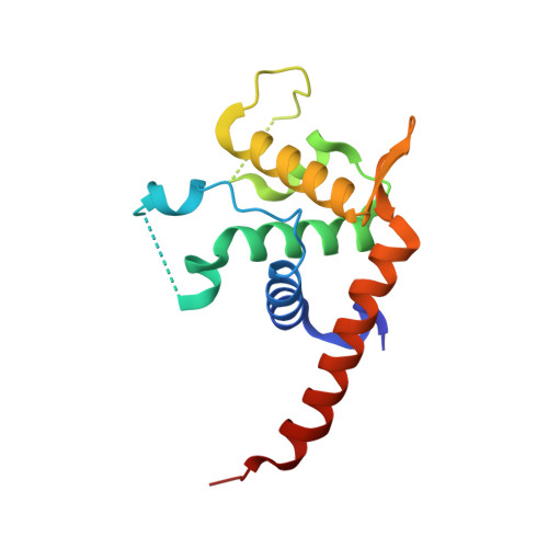
    Diamond-Blackfan anemia (DBA) is a rare congenital disease linked to mutations in the ribosomal protein genes rps19, rps24 and rps17. It belongs to the emerging class of ribosomal disorders. To understand the impact of DBA mutations on RPS19 function, we have solved the crystal structure of RPS19 from Pyrococcus abyssi. The protein forms a five alpha-helix bundle organized around a central amphipathic alpha-helix, which corresponds to the DBA mutation hot spot. From the structure, we classify DBA mutations relative to their respective impact on protein folding (class I) or on surface properties (class II). Class II mutations cluster into two conserved basic patches. In vivo analysis in yeast demonstrates an essential role for class II residues in the incorporation into pre-40S ribosomal particles. This data indicate that missense mutations in DBA primarily affect the capacity of the protein to be incorporated into pre-ribosomes, thus blocking maturation of the pre-40S particles.


  • Organizational Affiliation

    INSERM U869, Institut Européen de Chimie et Biologie, 2 rue Robert Escarpit Pessac, F-33607, France.


Macromolecules
Find similar proteins by:  (by identity cutoff)  |  3D Structure
Entity ID: 1
MoleculeChains Sequence LengthOrganismDetailsImage
RPS19E SSU RIBOSOMAL PROTEIN S19E150Pyrococcus abyssiMutation(s): 0 
UniProt
Find proteins for Q9V0G8 (Pyrococcus abyssi (strain GE5 / Orsay))
Explore Q9V0G8 
Go to UniProtKB:  Q9V0G8
Entity Groups  
Sequence Clusters30% Identity50% Identity70% Identity90% Identity95% Identity100% Identity
UniProt GroupQ9V0G8
Sequence Annotations
Expand
  • Reference Sequence
Small Molecules
Ligands 1 Unique
IDChains Name / Formula / InChI Key2D Diagram3D Interactions
CL
Query on CL

Download Ideal Coordinates CCD File 
B [auth A]CHLORIDE ION
Cl
VEXZGXHMUGYJMC-UHFFFAOYSA-M
Experimental Data & Validation

Experimental Data

  • Method: X-RAY DIFFRACTION
  • Resolution: 1.15 Å
  • R-Value Free: 0.155 
  • R-Value Work: 0.137 
  • R-Value Observed: 0.138 
  • Space Group: P 21 21 21
Unit Cell:
Length ( Å )Angle ( ˚ )
a = 32.7α = 90
b = 57.43β = 90
c = 80.2γ = 90
Software Package:
Software NamePurpose
REFMACrefinement
XDSdata reduction
XSCALEdata scaling
SHARPphasing

Structure Validation

View Full Validation Report


Entry History 

Deposition Data

Revision History  (Full details and data files)

  • Version 1.0: 2007-09-11
    Type: Initial release
  • Version 1.1: 2011-05-08
    Changes: Version format compliance
  • Version 1.2: 2011-07-13
    Changes: Version format compliance