2V2F

Crystal structure of PBP1a from drug-resistant strain 5204 from Streptococcus pneumoniae


Experimental Data Snapshot

  • Method: X-RAY DIFFRACTION
  • Resolution: 1.90 Å
  • R-Value Free: 0.246 
  • R-Value Work: 0.210 
  • R-Value Observed: 0.213 

wwPDB Validation   3D Report Full Report


This is version 1.3 of the entry. See complete history


Literature

Common Alterations in Pbp1A from Resistant Streptococcus Pneumoniae Decrease its Reactivity Toward {Beta}-Lactams: Structural Insights.

Job, V.Carapito, R.Vernet, T.Dessen, A.Zapun, A.

(2008) J Biol Chem 283: 4886

  • DOI: https://doi.org/10.1074/jbc.M706181200
  • Primary Citation of Related Structures:  
    2V2F

  • PubMed Abstract: 

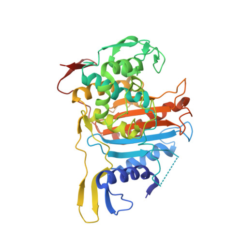
    The development of high level beta-lactam resistance in the pneumococcus requires the expression of an altered form of PBP1a, in addition to modified forms of PBP2b and PBP2x, which are necessary for the appearance of low levels of resistance. Here, we present the crystal structure of a soluble form of PBP1a from the highly resistant Streptococcus pneumoniae strain 5204 (minimal inhibitory concentration of cefotaxime is 12 mg.liter(-1)). Mutations T371A, which is adjacent to the catalytic nucleophile Ser370, and TSQF(574-577)NTGY, which lie in a loop bordering the active site cleft, were investigated by site-directed mutagenesis. The consequences of these substitutions on reaction kinetics with beta-lactams were probed in vitro, and their effect on resistance was measured in vivo. The results are interpreted in the framework of the crystal structure, which displays a narrower, discontinuous active site cavity, compared with that of PBP1a from the beta-lactam susceptible strain R6, as well as a reorientation of the catalytic Ser370.


  • Organizational Affiliation

    Laboratoire des Protéines Membranaires, Institut de Biologie Structurale Jean-Pierre Ebel, Université Joseph Fourier, UMR 5075-CNRS, CEA Grenoble, France.


Macromolecules

Find similar proteins by:  Sequence   |   3D Structure  

Entity ID: 1
MoleculeChains Sequence LengthOrganismDetailsImage
PENICILLIN BINDING PROTEIN 1A24Streptococcus pneumoniaeMutation(s): 0 
EC: 2.4.1.129
UniProt
Find proteins for Q9RET4 (Streptococcus pneumoniae)
Explore Q9RET4 
Go to UniProtKB:  Q9RET4
Entity Groups  
Sequence Clusters30% Identity50% Identity70% Identity90% Identity95% Identity100% Identity
UniProt GroupQ9RET4
Sequence Annotations
Expand
  • Reference Sequence
Find similar proteins by:  (by identity cutoff)  |  3D Structure
Entity ID: 2
MoleculeChains Sequence LengthOrganismDetailsImage
PENICILLIN BINDING PROTEIN 1AB [auth F]390Streptococcus pneumoniaeMutation(s): 0 
EC: 3.4.16.4
UniProt
Find proteins for Q9RET4 (Streptococcus pneumoniae)
Explore Q9RET4 
Go to UniProtKB:  Q9RET4
Entity Groups  
Sequence Clusters30% Identity50% Identity70% Identity90% Identity95% Identity100% Identity
UniProt GroupQ9RET4
Sequence Annotations
Expand
  • Reference Sequence
Small Molecules
Ligands 2 Unique
IDChains Name / Formula / InChI Key2D Diagram3D Interactions
MES
Query on MES

Download Ideal Coordinates CCD File 
D [auth F]2-(N-MORPHOLINO)-ETHANESULFONIC ACID
C6 H13 N O4 S
SXGZJKUKBWWHRA-UHFFFAOYSA-N
BA
Query on BA

Download Ideal Coordinates CCD File 
C [auth F]BARIUM ION
Ba
XDFCIPNJCBUZJN-UHFFFAOYSA-N
Experimental Data & Validation

Experimental Data

  • Method: X-RAY DIFFRACTION
  • Resolution: 1.90 Å
  • R-Value Free: 0.246 
  • R-Value Work: 0.210 
  • R-Value Observed: 0.213 
  • Space Group: C 1 2 1
Unit Cell:
Length ( Å )Angle ( ˚ )
a = 122.96α = 90
b = 67.03β = 100.89
c = 49.13γ = 90
Software Package:
Software NamePurpose
REFMACrefinement
XDSdata reduction
XSCALEdata scaling
AMoREphasing

Structure Validation

View Full Validation Report


Entry History 

Deposition Data

Revision History  (Full details and data files)

  • Version 1.0: 2007-12-25
    Type: Initial release
  • Version 1.1: 2011-05-08
    Changes: Version format compliance
  • Version 1.2: 2011-07-13
    Changes: Version format compliance
  • Version 1.3: 2023-12-13
    Changes: Data collection, Database references, Derived calculations, Refinement description