Structural basis for recruitment of tandem hotdog domains in acyl-CoA thioesterase 7 and its role in inflammation.
Forwood, J.K., Thakur, A.S., Guncar, G., Marfori, M., Mouradov, D., Meng, W., Robinson, J., Huber, T., Kellie, S., Martin, J.L., Hume, D.A., Kobe, B.(2007) Proc Natl Acad Sci U S A 104: 10382-10387
- PubMed: 17563367
- DOI: https://doi.org/10.1073/pnas.0700974104
- Primary Citation of Related Structures:
2Q2B, 2V1O - PubMed Abstract:
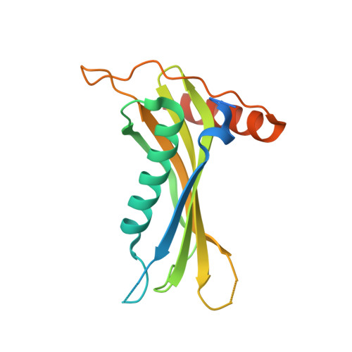
Acyl-CoA thioesterases (Acots) catalyze the hydrolysis of fatty acyl-CoA to free fatty acid and CoA and thereby regulate lipid metabolism and cellular signaling. We present a comprehensive structural and functional characterization of mouse acyl-CoA thioesterase 7 (Acot7). Whereas prokaryotic homologues possess a single thioesterase domain, mammalian Acot7 contains a pair of domains in tandem. We determined the crystal structures of both the N- and C-terminal domains of the mouse enzyme, and inferred the structure of the full-length enzyme using a combination of chemical cross-linking, mass spectrometry, and molecular modeling. The quaternary arrangement in Acot7 features a trimer of hotdog fold dimers. Both domains of Acot7 are required for activity, but only one of two possible active sites in the dimer is functional. Asn-24 and Asp-213 (from N- and C-domains, respectively) were identified as the catalytic residues through site-directed mutagenesis. An enzyme with higher activity than wild-type Acot7 was obtained by mutating the residues in the nonfunctional active site. Recombinant Acot7 was shown to have the highest activity toward arachidonoyl-CoA, suggesting a function in eicosanoid metabolism. In line with the proposal, Acot7 was shown to be highly expressed in macrophages and up-regulated by lipopolysaccharide. Overexpression of Acot7 in a macrophage cell line modified the production of prostaglandins D2 and E2. Together, the results link the molecular and cellular functions of Acot7 and identify the enzyme as a candidate drug target in inflammatory disease.
Organizational Affiliation:
School of Molecular and Microbial Sciences, University of Queensland, Brisbane, Queensland 4072, Australia. jforwood@csu.edu.au