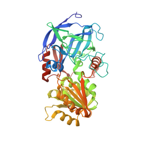
Refined structure of Cu-substituted alcohol dehydrogenase at 2.1 A resolution.
Al-Karadaghi, S., Cedergren-Zeppezauer, E.S., Dauter, Z., Wilson, K.S.(1995) Acta Crystallogr D Biol Crystallogr 51: 805-813
- PubMed: 15299812
- DOI: https://doi.org/10.1107/S090744499500045X
- Primary Citation of Related Structures:
2OXI - PubMed Abstract:
Liver alcohol dehydrogenase (LADH) is a Zn(II)-dependent dimeric enzyme. LADH with the active-site Zn(II) substituted by Cu(II) resembles blue (type I) copper proteins by its spectroscopic characteristics. In this work we present the X-ray structure of the active site Cu(II)-substituted LADH complex with NADH and dimethyl sulfoxide (DMSO). The structure was solved by molecular replacement. The space group is P2(1) with cell dimensions a = 44.4, b = 180.6, c = 50.8 A and beta = 108 degrees. There is one dimer of the enzyme in the asymmetric unit. The refinement was carried out to a crystallographic R-factor of 16.1% for 41 119 unique reflections in the resolution range 12.0 to 2.1 A. The coordination geometry of Cu(II) in LADH is compared with the active-site metal coordination in the Zn-LADH-NADH-DMSO complex and blue-copper proteins. The distances from the metal to the protein ligands (Cys46, His67 and Cys174) are similar for the Zn(II) and Cu(II) ions. The distances of the O atom of the inhibitor DMSO to the Cu(II) ion in the two subunits of the dimer are 3.19 and 3.45 A. These are considerably longer than the corresponding distances for the Zn(II) enzyme, 2.19 and 2.15 A. The Cu(II) ion is positioned nearly in the plane of the three protein ligands (NS(2)) with a geometry similar to the trigonal arrangement of the three strongly bound ligands (N(2)S) in blue-copper proteins. This coordination probably accounts for the similarity of the spectral characteristics of Cu(II)-LADH and type I copper proteins.
Organizational Affiliation:
Department of Structural Chemistry, Arrhenius Laboratories for Natural Sciences, Stockholm University, Sweden.