2O9O

Crystal Structure of the buffalo Secretory Signalling Glycoprotein at 2.8 A resolution


Experimental Data Snapshot

  • Method: X-RAY DIFFRACTION
  • Resolution: 2.80 Å
  • R-Value Free: 0.236 
  • R-Value Work: 0.186 
  • R-Value Observed: 0.188 

wwPDB Validation   3D Report Full Report


This is version 2.1 of the entry. See complete history


Literature

Structure of the buffalo secretory signalling glycoprotein at 2.8 A resolution

Ethayathulla, A.S.Srivastava, D.B.Kumar, J.Saravanan, K.Bilgrami, S.Sharma, S.Kaur, P.Srinivasan, A.Singh, T.P.

(2007) Acta Crystallogr Sect F Struct Biol Cryst Commun 63: 258-265

  • DOI: https://doi.org/10.1107/S1744309107010445
  • Primary Citation of Related Structures:  
    2O9O

  • PubMed Abstract: 

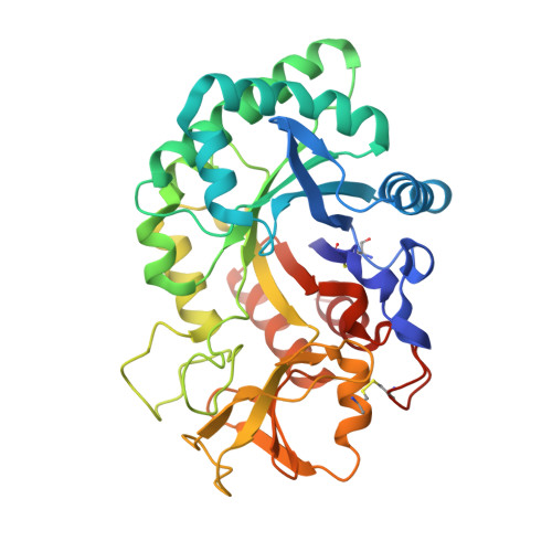
    The crystal structure of a 40 kDa signalling glycoprotein from buffalo (SPB-40) has been determined at 2.8 A resolution. SPB-40 acts as a protective signalling factor by binding to viable cells during the early phase of involution, during which extensive tissue remodelling occurs. It was isolated from the dry secretions of Murrah buffalo. It was purified and crystallized using the hanging-drop vapour-diffusion method with 19% ethanol as the precipitant. The protein was also cloned and its complete nucleotide and amino-acid sequences were determined. When compared with the sequences of other members of the family, the sequence of SPB-40 revealed two very important mutations in the sugar-binding region, in which Tyr120 changed to Trp120 and Glu269 changed to Trp269. The structure showed a significant distortion in the shape of the sugar-binding groove. The water structure in the groove is also drastically altered. The folding of the protein chain in the flexible region comprising segments His188-His197, Phe202-Arg212 and Tyr244-Pro260 shows large variations when compared with other proteins of the family.


  • Organizational Affiliation

    Department of Biophysics, All India Institute of Medical Sciences, New Delhi 110029, India.


Macromolecules
Find similar proteins by:  (by identity cutoff)  |  3D Structure
Entity ID: 1
MoleculeChains Sequence LengthOrganismDetailsImage
Chitinase-3-like protein 1361Bubalus bubalisMutation(s): 0 
UniProt
Find proteins for Q7YS85 (Bubalus bubalis)
Explore Q7YS85 
Go to UniProtKB:  Q7YS85
Entity Groups  
Sequence Clusters30% Identity50% Identity70% Identity90% Identity95% Identity100% Identity
UniProt GroupQ7YS85
Sequence Annotations
Expand
  • Reference Sequence
Oligosaccharides

Help

Entity ID: 2
MoleculeChains Length2D Diagram Glycosylation3D Interactions
alpha-D-mannopyranose-(1-3)-[alpha-D-mannopyranose-(1-6)]alpha-D-mannopyranose-(1-4)-2-acetamido-2-deoxy-beta-D-glucopyranose-(1-4)-2-acetamido-2-deoxy-beta-D-glucopyranose
B
5N-Glycosylation
Glycosylation Resources
GlyTouCan:  G21381MC
GlyCosmos:  G21381MC
GlyGen:  G21381MC
Experimental Data & Validation

Experimental Data

  • Method: X-RAY DIFFRACTION
  • Resolution: 2.80 Å
  • R-Value Free: 0.236 
  • R-Value Work: 0.186 
  • R-Value Observed: 0.188 
  • Space Group: P 21 21 21
Unit Cell:
Length ( Å )Angle ( ˚ )
a = 63.1α = 90
b = 66.859β = 90
c = 108.543γ = 90
Software Package:
Software NamePurpose
REFMACrefinement
MAR345data collection
DENZOdata reduction
SCALEPACKdata scaling
AMoREphasing

Structure Validation

View Full Validation Report


Entry History 

Deposition Data

Revision History  (Full details and data files)

  • Version 1.0: 2007-01-02
    Type: Initial release
  • Version 1.1: 2008-05-01
    Changes: Version format compliance
  • Version 1.2: 2011-07-13
    Changes: Non-polymer description, Version format compliance
  • Version 1.3: 2017-10-18
    Changes: Refinement description
  • Version 2.0: 2020-07-29
    Type: Remediation
    Reason: Carbohydrate remediation
    Changes: Advisory, Atomic model, Data collection, Derived calculations, Structure summary
  • Version 2.1: 2023-10-25
    Changes: Data collection, Database references, Refinement description, Structure summary