2IBB

Crystal Structure of the First and Second FNIII Domains of Ihog


Experimental Data Snapshot

  • Method: X-RAY DIFFRACTION
  • Resolution: 2.40 Å
  • R-Value Free: 0.275 
  • R-Value Work: 0.244 

wwPDB Validation   3D Report Full Report


This is version 1.4 of the entry. See complete history


Literature

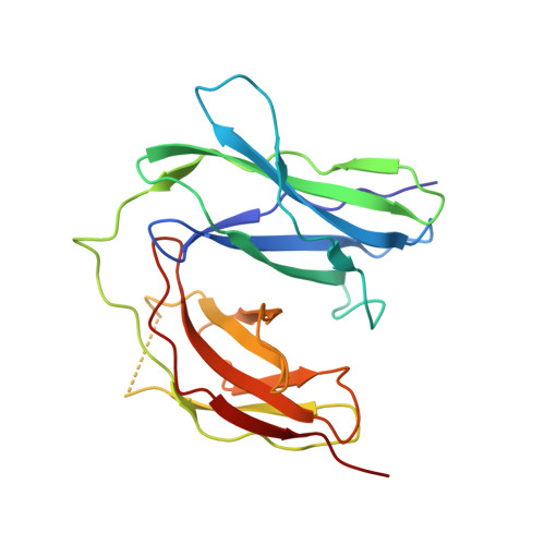
Structure of a heparin-dependent complex of Hedgehog and Ihog.

McLellan, J.S.Yao, S.Zheng, X.Geisbrecht, B.V.Ghirlando, R.Beachy, P.A.Leahy, D.J.

(2006) Proc Natl Acad Sci U S A 103: 17208-17213

  • DOI: https://doi.org/10.1073/pnas.0606738103
  • Primary Citation of Related Structures:  
    2IBB, 2IBG, 2IC2

  • PubMed Abstract: 

    Hedgehog (Hh) signaling molecules mediate key tissue-patterning events during animal development, and inappropriate activation of Hh signaling in adults has been associated with human cancers. Recently, a conserved family of type I integral membrane proteins required for normal response to the Hh signal was discovered. One member of this family, Ihog (interference hedgehog), functions upstream or at the level of Patched (Ptc), but how Ihog participates in Hh signaling remains unclear. Here, we show that heparin binding induces Ihog dimerization and is required to mediate high-affinity interactions between Ihog and Hh. We also present crystal structures of a Hh-binding fragment of Ihog, both alone and complexed with Hh. Heparin is not well ordered in these structures, but a basic cleft in the first FNIII domain of Ihog (IhogFn1) is shown by mutagenesis to mediate heparin binding. These results establish that Hh directly binds Ihog and provide the first demonstration of a specific role for heparin in Hh responsiveness.


  • Organizational Affiliation

    Department of Biophysics and Biophysical Chemistry, Johns Hopkins University School of Medicine, Baltimore, MD 21205, USA.


Macromolecules
Find similar proteins by:  (by identity cutoff)  |  3D Structure
Entity ID: 1
MoleculeChains Sequence LengthOrganismDetailsImage
CG9211-PA213Drosophila melanogasterMutation(s): 0 
Gene Names: iHogCG9211
UniProt
Find proteins for Q9VM64 (Drosophila melanogaster)
Explore Q9VM64 
Go to UniProtKB:  Q9VM64
Entity Groups  
Sequence Clusters30% Identity50% Identity70% Identity90% Identity95% Identity100% Identity
UniProt GroupQ9VM64
Sequence Annotations
Expand
  • Reference Sequence
Experimental Data & Validation

Experimental Data

  • Method: X-RAY DIFFRACTION
  • Resolution: 2.40 Å
  • R-Value Free: 0.275 
  • R-Value Work: 0.244 
  • Space Group: P 21 21 21
Unit Cell:
Length ( Å )Angle ( ˚ )
a = 39.38α = 90
b = 65.24β = 90
c = 84.21γ = 90
Software Package:
Software NamePurpose
DENZOdata reduction
SCALEPACKdata scaling
CNSrefinement
PDB_EXTRACTdata extraction
HKL-2000data reduction
HKL-2000data scaling
AMoREphasing

Structure Validation

View Full Validation Report


Entry History 

Deposition Data

Revision History  (Full details and data files)

  • Version 1.0: 2006-10-24
    Type: Initial release
  • Version 1.1: 2008-05-01
    Changes: Version format compliance
  • Version 1.2: 2011-07-13
    Changes: Version format compliance
  • Version 1.3: 2017-10-18
    Changes: Refinement description
  • Version 1.4: 2023-08-30
    Changes: Data collection, Database references, Derived calculations, Refinement description