Structural basis of CoA recognition by the Pyrococcus single-domain CoA-binding proteins.
Hiyama, T.B., Zhao, M., Kitago, Y., Yao, M., Sekine, S., Terada, T., Kuroishi, C., Liu, Z.J., Rose, J.P., Kuramitsu, S., Shirouzu, M., Watanabe, N., Yokoyama, S., Tanaka, I., Wang, B.C.(2006) J Struct Funct Genomics 7: 119-129
- PubMed: 17342453
- DOI: https://doi.org/10.1007/s10969-007-9015-6
- Primary Citation of Related Structures:
2D59, 2D5A - PubMed Abstract:
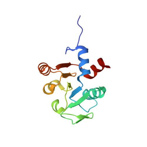
The single-domain coenzyme A (CoA)-binding protein is conserved in bacteria, archaea, and a few eukaryal taxa. It consists of a Rossmann-fold domain, belonging to the FAD/NAD(P)-binding ;superfamily. The crystal structure of the Thermus thermophilus single-domain CoA-binding protein, TTHA1899, has been determined and it has been demonstrated, by isothermal titration calorimetry, that the protein interacts with CoA [Wada T. et al. Acta Crystallogr D Biol Crystallogr 59 (2003) 1213]. In the present study, we determined the crystal structures of an orthologous protein from the archaeon Pyrococcus horikoshii (PH1109), alone and complexed with CoA, at 1.65 A and 1.70 A resolutions, respectively, and that of P. furiosus protein (PF0725) in the CoA-bound form at 1.70 A. The CoA-bound structures are very similar to each other, revealing that the Pyrococcus proteins bind CoA in a 1:1 stoichiometry. Five loop-containing regions form the CoA-binding groove, to which the CoA molecule is docked. A comparison of the structures and the sequences of the Pyrococcus proteins with those of the T. theromphilus orthologue TTHA1899 indicated that archaeal and bacterial single-domain CoA-binding proteins share the same CoA-binding mode. Nevertheless, many of the peripheral residues involved in the hydrogen-bonding/electrostatic interactions with CoA are not strictly conserved in the family. The CoA interaction of the single-domain CoA-binding proteins is significantly different and much more extensive than that of the multi-subunit/multi-domain CoA-binding protein succinyl-CoA synthetase.
Organizational Affiliation:
Department of Biophysics and Biochemistry, Graduate School of Science, The University of Tokyo, 7-3-1 Hongo, Bunkyo-ku, Tokyo 113-0033, Japan.