2BEH

Crystal structure of antithrombin variant S137A/V317C/T401C with plasma latent antithrombin


Experimental Data Snapshot

  • Method: X-RAY DIFFRACTION
  • Resolution: 2.70 Å
  • R-Value Free: 0.265 
  • R-Value Work: 0.219 
  • R-Value Observed: 0.219 

wwPDB Validation   3D Report Full Report


Ligand Structure Quality Assessment 


This is version 2.1 of the entry. See complete history


Literature

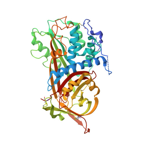
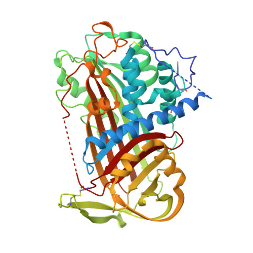
Crystal structure of monomeric native antithrombin reveals a novel reactive center loop conformation.

Johnson, D.J.Langdown, J.Li, W.Luis, S.A.Baglin, T.P.Huntington, J.A.

(2006) J Biol Chem 281: 35478-35486

  • DOI: https://doi.org/10.1074/jbc.M607204200
  • Primary Citation of Related Structures:  
    1T1F, 2B5T, 2BEH

  • PubMed Abstract: 

    The poor inhibitory activity of circulating antithrombin (AT) is critical to the formation of blood clots at sites of vascular damage. AT becomes an efficient inhibitor of the coagulation proteases only after binding to a specific heparin pentasaccharide, which alters the conformation of the reactive center loop (RCL). The molecular basis of this activation event lies at the heart of the regulation of hemostasis and accounts for the anticoagulant properties of the low molecular weight heparins. Although several structures of AT have been solved, the conformation of the RCL in native AT remains unknown because of the obligate crystal contact between the RCL of native AT and its latent counterpart. Here we report the crystallographic structure of a variant of AT in its monomeric native state. The RCL shifted approximately 20 A, and a salt bridge was observed between the P1 residue (Arg-393) and Glu-237. This contact explains the effect of mutations at the P1 position on the affinity of AT for heparin and also the properties of AT-Truro (E237K). The relevance of the observed conformation was verified through mutagenesis studies and by solving structures of the same variant in different crystal forms. We conclude that the poor inhibitory activity of the circulating form of AT is partially conferred by intramolecular contacts that restrain the RCL, orient the P1 residue away from attacking proteases, and additionally block the exosite utilized in protease recognition.


  • Organizational Affiliation

    Department of Haematology, Division of Structural Medicine, Thrombosis Research Unit, Cambridge Institute for Medical Research, University of Cambridge, Wellcome Trust/MRC Building, Hills Road, Cambridge CB2 2XY, United Kingdom.


Macromolecules
Find similar proteins by:  (by identity cutoff)  |  3D Structure
Entity ID: 1
MoleculeChains Sequence LengthOrganismDetailsImage
Antithrombin-IIIA [auth I]432Homo sapiensMutation(s): 3 
Gene Names: SERPINC1AT3
UniProt & NIH Common Fund Data Resources
Find proteins for P01008 (Homo sapiens)
Explore P01008 
Go to UniProtKB:  P01008
PHAROS:  P01008
GTEx:  ENSG00000117601 
Entity Groups  
Sequence Clusters30% Identity50% Identity70% Identity90% Identity95% Identity100% Identity
UniProt GroupP01008
Sequence Annotations
Expand
  • Reference Sequence
Find similar proteins by:  (by identity cutoff)  |  3D Structure
Entity ID: 2
MoleculeChains Sequence LengthOrganismDetailsImage
Antithrombin-IIIB [auth L]432Homo sapiensMutation(s): 0 
UniProt & NIH Common Fund Data Resources
Find proteins for P01008 (Homo sapiens)
Explore P01008 
Go to UniProtKB:  P01008
PHAROS:  P01008
GTEx:  ENSG00000117601 
Entity Groups  
Sequence Clusters30% Identity50% Identity70% Identity90% Identity95% Identity100% Identity
UniProt GroupP01008
Sequence Annotations
Expand
  • Reference Sequence
Oligosaccharides

Help

Entity ID: 3
MoleculeChains Length2D Diagram Glycosylation3D Interactions
2-acetamido-2-deoxy-beta-D-glucopyranose-(1-4)-2-acetamido-2-deoxy-beta-D-glucopyranoseC [auth A],
E [auth C]
2N-Glycosylation
Glycosylation Resources
GlyTouCan:  G42666HT
GlyCosmos:  G42666HT
GlyGen:  G42666HT
Entity ID: 4
MoleculeChains Length2D Diagram Glycosylation3D Interactions
alpha-D-mannopyranose-(1-4)-2-acetamido-2-deoxy-beta-D-glucopyranose-(1-4)-2-acetamido-2-deoxy-beta-D-glucopyranoseD [auth B]3N-Glycosylation
Glycosylation Resources
GlyTouCan:  G62182OO
GlyCosmos:  G62182OO
GlyGen:  G62182OO
Experimental Data & Validation

Experimental Data

  • Method: X-RAY DIFFRACTION
  • Resolution: 2.70 Å
  • R-Value Free: 0.265 
  • R-Value Work: 0.219 
  • R-Value Observed: 0.219 
  • Space Group: P 1 21 1
Unit Cell:
Length ( Å )Angle ( ˚ )
a = 68.073α = 90
b = 97.274β = 105.03
c = 87.388γ = 90
Software Package:
Software NamePurpose
CNSrefinement
MOSFLMdata reduction
CCP4data scaling
MOLREPphasing

Structure Validation

View Full Validation Report


Ligand Structure Quality Assessment 


Entry History 

Deposition Data

Revision History  (Full details and data files)

  • Version 1.0: 2005-11-01
    Type: Initial release
  • Version 1.1: 2008-05-01
    Changes: Version format compliance
  • Version 1.2: 2011-07-13
    Changes: Non-polymer description, Version format compliance
  • Version 2.0: 2020-07-29
    Type: Remediation
    Reason: Carbohydrate remediation
    Changes: Advisory, Atomic model, Data collection, Derived calculations, Non-polymer description, Structure summary
  • Version 2.1: 2021-10-20
    Changes: Database references, Structure summary