2A8X

Crystal Structure of Lipoamide Dehydrogenase from Mycobacterium tuberculosis


Experimental Data Snapshot

  • Method: X-RAY DIFFRACTION
  • Resolution: 2.40 Å
  • R-Value Free: 0.246 
  • R-Value Work: 0.199 
  • R-Value Observed: 0.199 

wwPDB Validation   3D Report Full Report


Ligand Structure Quality Assessment 


This is version 1.3 of the entry. See complete history


Literature

Crystal structure and functional analysis of lipoamide dehydrogenase from Mycobacterium tuberculosis

Rajashankar, K.R.Bryk, R.Kniewel, R.Buglino, J.A.Nathan, C.F.Lima, C.D.

(2005) J Biol Chem 280: 33977-33983

  • DOI: https://doi.org/10.1074/jbc.M507466200
  • Primary Citation of Related Structures:  
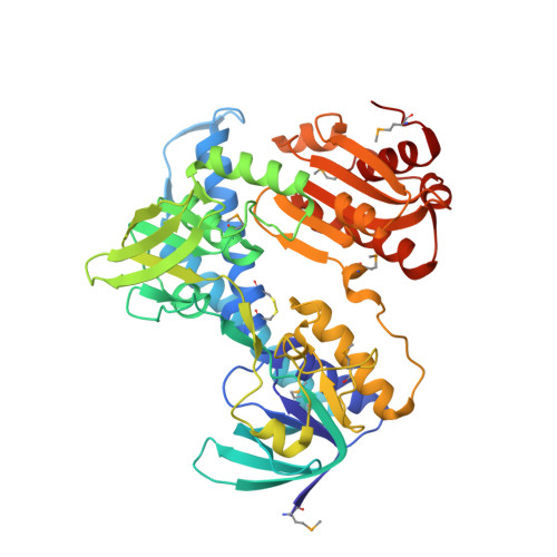
    2A8X

  • PubMed Abstract: 

    We report the 2.4 A crystal structure for lipoamide dehydrogenase encoded by lpdC from Mycobacterium tuberculosis. Based on the Lpd structure and sequence alignment between bacterial and eukaryotic Lpd sequences, we generated single point mutations in Lpd and assayed the resulting proteins for their ability to catalyze lipoamide reduction/oxidation alone and in complex with other proteins that participate in pyruvate dehydrogenase and peroxidase activities. The results suggest that amino acid residues conserved in mycobacterial species but not conserved in eukaryotic Lpd family members modulate either or both activities and include Arg-93, His-98, Lys-103, and His-386. In addition, Arg-93 and His-386 are involved in forming both "open" and "closed" active site conformations, suggesting that these residues play a role in dynamically regulating Lpd function. Taken together, these data suggest protein surfaces that should be considered while developing strategies for inhibiting this enzyme.


  • Organizational Affiliation

    Structural Biology Program, Sloan-Kettering Institute, New York, New York 10021, USA.


Macromolecules
Find similar proteins by:  (by identity cutoff)  |  3D Structure
Entity ID: 1
MoleculeChains Sequence LengthOrganismDetailsImage
Dihydrolipoyl dehydrogenase
A, B
464Mycobacterium tuberculosisMutation(s): 7 
Gene Names: Rv0462MT0478lpdC
EC: 1.8.1.4
UniProt
Find proteins for P9WHH9 (Mycobacterium tuberculosis (strain ATCC 25618 / H37Rv))
Explore P9WHH9 
Go to UniProtKB:  P9WHH9
Entity Groups  
Sequence Clusters30% Identity50% Identity70% Identity90% Identity95% Identity100% Identity
UniProt GroupP9WHH9
Sequence Annotations
Expand
  • Reference Sequence
Small Molecules
Modified Residues  1 Unique
IDChains TypeFormula2D DiagramParent
MSE
Query on MSE
A, B
L-PEPTIDE LINKINGC5 H11 N O2 SeMET
Experimental Data & Validation

Experimental Data

  • Method: X-RAY DIFFRACTION
  • Resolution: 2.40 Å
  • R-Value Free: 0.246 
  • R-Value Work: 0.199 
  • R-Value Observed: 0.199 
  • Space Group: P 21 21 21
Unit Cell:
Length ( Å )Angle ( ˚ )
a = 83.582α = 90
b = 96.677β = 90
c = 122.889γ = 90
Software Package:
Software NamePurpose
DENZOdata reduction
SCALEPACKdata scaling
SOLVEphasing
CNSrefinement

Structure Validation

View Full Validation Report


Ligand Structure Quality Assessment 


Entry History 

Deposition Data

Revision History  (Full details and data files)

  • Version 1.0: 2005-08-16
    Type: Initial release
  • Version 1.1: 2008-04-30
    Changes: Version format compliance
  • Version 1.2: 2011-07-13
    Changes: Version format compliance
  • Version 1.3: 2024-04-03
    Changes: Data collection, Database references, Derived calculations, Refinement description, Structure summary