1YT5

Crystal structure of NAD kinase from Thermotoga maritima


Experimental Data Snapshot

  • Method: X-RAY DIFFRACTION
  • Resolution: 2.30 Å
  • R-Value Free: 0.280 
  • R-Value Work: 0.213 
  • R-Value Observed: 0.217 

wwPDB Validation   3D Report Full Report


This is version 1.4 of the entry. See complete history


Literature

Structure of a NAD kinase from Thermotoga maritima at 2.3 A resolution.

Oganesyan, V.Huang, C.Adams, P.D.Jancarik, J.Yokota, H.A.Kim, R.Kim, S.H.

(2005) Acta Crystallogr Sect F Struct Biol Cryst Commun 61: 640-646

  • DOI: https://doi.org/10.1107/S1744309105019780
  • Primary Citation of Related Structures:  
    1YT5

  • PubMed Abstract: 

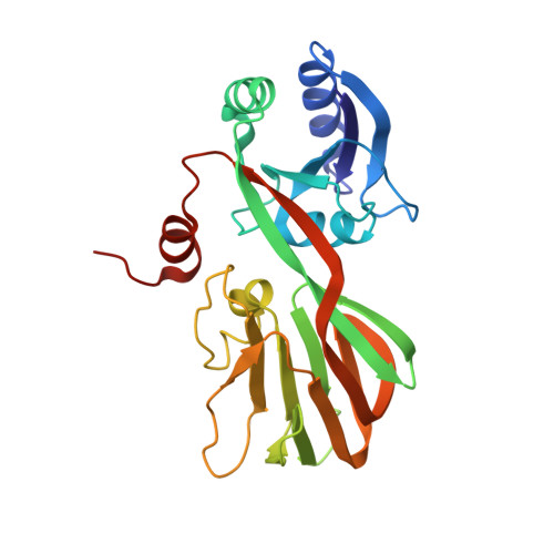
    NAD kinase is the only known enzyme that catalyzes the formation of NADP, a coenzyme involved in most anabolic reactions and in the antioxidant defense system. Despite its importance, very little is known regarding the mechanism of catalysis and only recently have several NAD kinase structures been deposited in the PDB. Here, an independent investigation of the crystal structure of inorganic polyphosphate/ATP-NAD kinase, PPNK_THEMA, a protein from Thermotoga maritima, is reported at a resolution of 2.3 A. The crystal structure was solved using single-wavelength anomalous diffraction (SAD) data collected at the Se absorption-peak wavelength in a state in which no cofactors or substrates were bound. It revealed that the 258-amino-acid protein is folded into two distinct domains, similar to recently reported NAD kinases. The N-terminal alpha/beta-domain spans the first 100 amino acids and the last 30 amino acids of the polypeptide and has several topological matches in the PDB, whereas the other domain, which spans the middle 130 residues, adopts a unique beta-sandwich architecture and only appreciably matches the recently deposited PDB structures of NAD kinases.


  • Organizational Affiliation

    Structural Genomics Center, Physical Biosciences Division, Lawrence Berkeley National Laboratory, Berkeley, California 94720, USA.


Macromolecules
Find similar proteins by:  (by identity cutoff)  |  3D Structure
Entity ID: 1
MoleculeChains Sequence LengthOrganismDetailsImage
inorganic polyphosphate/ATP-NAD kinase
A, B, C, D
258Thermotoga maritimaMutation(s): 0 
Gene Names: ppnK
EC: 2.7.1.23
UniProt
Find proteins for Q9X255 (Thermotoga maritima (strain ATCC 43589 / DSM 3109 / JCM 10099 / NBRC 100826 / MSB8))
Explore Q9X255 
Go to UniProtKB:  Q9X255
Entity Groups  
Sequence Clusters30% Identity50% Identity70% Identity90% Identity95% Identity100% Identity
UniProt GroupQ9X255
Sequence Annotations
Expand
  • Reference Sequence
Small Molecules
Ligands 1 Unique
IDChains Name / Formula / InChI Key2D Diagram3D Interactions
SO4
Query on SO4

Download Ideal Coordinates CCD File 
E [auth A]
F [auth A]
G [auth A]
H [auth A]
I [auth A]
E [auth A],
F [auth A],
G [auth A],
H [auth A],
I [auth A],
J [auth A],
K [auth B],
L [auth B],
M [auth B],
N [auth C],
O [auth C],
P [auth D],
Q [auth D],
R [auth D],
S [auth D],
T [auth D],
U [auth D],
V [auth D]
SULFATE ION
O4 S
QAOWNCQODCNURD-UHFFFAOYSA-L
Experimental Data & Validation

Experimental Data

  • Method: X-RAY DIFFRACTION
  • Resolution: 2.30 Å
  • R-Value Free: 0.280 
  • R-Value Work: 0.213 
  • R-Value Observed: 0.217 
  • Space Group: P 21 21 2
Unit Cell:
Length ( Å )Angle ( ˚ )
a = 131.448α = 90
b = 137.153β = 90
c = 58.253γ = 90
Software Package:
Software NamePurpose
HKL-2000data collection
SCALEPACKdata scaling
PHENIXmodel building
SOLVEphasing
REFMACrefinement
HKL-2000data reduction
PHENIXphasing

Structure Validation

View Full Validation Report


Entry History 

Deposition Data

Revision History  (Full details and data files)

  • Version 1.0: 2005-04-05
    Type: Initial release
  • Version 1.1: 2008-04-30
    Changes: Version format compliance
  • Version 1.2: 2011-07-13
    Changes: Version format compliance
  • Version 1.3: 2017-10-11
    Changes: Refinement description
  • Version 1.4: 2024-02-14
    Changes: Data collection, Database references, Derived calculations