1YD6

Crystal structure of the GIY-YIG N-terminal endonuclease domain of UvrC from Bacillus caldotenax


Experimental Data Snapshot

  • Method: X-RAY DIFFRACTION
  • Resolution: 2.00 Å
  • R-Value Free: 0.252 
  • R-Value Work: 0.202 
  • R-Value Observed: 0.205 

wwPDB Validation   3D Report Full Report


This is version 1.3 of the entry. See complete history


Literature

Structural insights into the first incision reaction during nucleotide excision repair

Truglio, J.J.Rhau, B.Croteau, D.L.Wang, L.Skorvaga, M.Karakas, E.Dellavecchia, M.J.Wang, H.Van Houten, B.Kisker, C.

(2005) EMBO J 24: 885-894

  • DOI: https://doi.org/10.1038/sj.emboj.7600568
  • Primary Citation of Related Structures:  
    1YCZ, 1YD0, 1YD1, 1YD2, 1YD3, 1YD4, 1YD5, 1YD6

  • PubMed Abstract: 

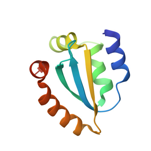
    Nucleotide excision repair is a highly conserved DNA repair mechanism present in all kingdoms of life. The incision reaction is a critical step for damage removal and is accomplished by the UvrC protein in eubacteria. No structural information is so far available for the 3' incision reaction. Here we report the crystal structure of the N-terminal catalytic domain of UvrC at 1.5 A resolution, which catalyzes the 3' incision reaction and shares homology with the catalytic domain of the GIY-YIG family of intron-encoded homing endonucleases. The structure reveals a patch of highly conserved residues surrounding a catalytic magnesium-water cluster, suggesting that the metal binding site is an essential feature of UvrC and all GIY-YIG endonuclease domains. Structural and biochemical data strongly suggest that the N-terminal endonuclease domain of UvrC utilizes a novel one-metal mechanism to cleave the phosphodiester bond.


  • Organizational Affiliation

    Department of Pharmacological Sciences, State University of New York at Stony Brook, Stony Brook, NY, USA.


Macromolecules
Find similar proteins by:  (by identity cutoff)  |  3D Structure
Entity ID: 1
MoleculeChains Sequence LengthOrganismDetailsImage
UvrC
A, B, C, D
99[Bacillus] caldotenaxMutation(s): 0 
UniProt
Find proteins for Q5KWH6 (Geobacillus kaustophilus (strain HTA426))
Explore Q5KWH6 
Go to UniProtKB:  Q5KWH6
Entity Groups  
Sequence Clusters30% Identity50% Identity70% Identity90% Identity95% Identity100% Identity
UniProt GroupQ5KWH6
Sequence Annotations
Expand
  • Reference Sequence
Experimental Data & Validation

Experimental Data

  • Method: X-RAY DIFFRACTION
  • Resolution: 2.00 Å
  • R-Value Free: 0.252 
  • R-Value Work: 0.202 
  • R-Value Observed: 0.205 
  • Space Group: C 1 2 1
Unit Cell:
Length ( Å )Angle ( ˚ )
a = 86.487α = 90
b = 86.66β = 120.16
c = 67.862γ = 90
Software Package:
Software NamePurpose
REFMACrefinement
DENZOdata reduction
SCALEPACKdata scaling
SOLVEphasing

Structure Validation

View Full Validation Report


Entry History 

Deposition Data

Revision History  (Full details and data files)

  • Version 1.0: 2005-03-01
    Type: Initial release
  • Version 1.1: 2008-04-30
    Changes: Version format compliance
  • Version 1.2: 2011-07-13
    Changes: Version format compliance
  • Version 1.3: 2024-03-13
    Changes: Data collection, Database references, Derived calculations