1XW4

Crystal Structure of Human Sulfiredoxin (Srx) in Complex with ADP


Experimental Data Snapshot

  • Method: X-RAY DIFFRACTION
  • Resolution: 2.00 Å
  • R-Value Free: 0.271 
  • R-Value Work: 0.218 
  • R-Value Observed: 0.221 

wwPDB Validation   3D Report Full Report


Ligand Structure Quality Assessment 


This is version 1.4 of the entry. See complete history


Literature

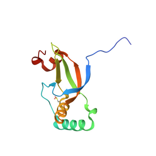
Structural basis for the retroreduction of inactivated peroxiredoxins by human sulfiredoxin.

Jonsson, T.J.Murray, M.S.Johnson, L.C.Poole, L.B.Lowther, W.T.

(2005) Biochemistry 44: 8634-8642

  • DOI: https://doi.org/10.1021/bi050131i
  • Primary Citation of Related Structures:  
    1XW3, 1XW4

  • PubMed Abstract: 

    Sufiredoxins (Srx) repair the inactivated forms of typical two-Cys peroxiredoxins (Prx) implicated in hydrogen peroxide-mediated cell signaling. The reduction of the cysteine sulfinic acid moiety within the active site of the Prx by Srx involves novel sulfur chemistry and the use of ATP and Mg(2+). The 1.65 A crystal structure of human Srx (hSrx) exhibits a new protein fold and a unique nucleotide binding motif containing the Gly98-Cys99-His100-Arg101 sequence at the N-terminus of an alpha-helix. HPLC analysis of the reaction products has confirmed that the site of ATP cleavage is between the beta- and gamma-phosphate groups. Cys99 and the gamma-phosphate of ATP, modeled within the active site of the 2.0 A ADP product complex structure, are adjacent to large surface depressions containing additional conserved residues. These features and the necessity for significant remodeling of the Prx structure suggest that the interactions between hSrx and typical two-Cys Prxs are specific. Moreover, the concave shape of the hSrx active site surface appears to be ideally suited to interacting with the convex surface of the toroidal Prx decamer.


  • Organizational Affiliation

    Center for Structural Biology, Department of Biochemistry, Wake Forest University School of Medicine, Medical Center Boulevard, Winston-Salem, North Carolina 27157, USA.


Macromolecules
Find similar proteins by:  (by identity cutoff)  |  3D Structure
Entity ID: 1
MoleculeChains Sequence LengthOrganismDetailsImage
SulfiredoxinA [auth X]110Homo sapiensMutation(s): 1 
UniProt & NIH Common Fund Data Resources
Find proteins for Q9BYN0 (Homo sapiens)
Explore Q9BYN0 
Go to UniProtKB:  Q9BYN0
PHAROS:  Q9BYN0
GTEx:  ENSG00000271303 
Entity Groups  
Sequence Clusters30% Identity50% Identity70% Identity90% Identity95% Identity100% Identity
UniProt GroupQ9BYN0
Sequence Annotations
Expand
  • Reference Sequence
Small Molecules
Ligands 1 Unique
IDChains Name / Formula / InChI Key2D Diagram3D Interactions
ADP
Query on ADP

Download Ideal Coordinates CCD File 
B [auth X]ADENOSINE-5'-DIPHOSPHATE
C10 H15 N5 O10 P2
XTWYTFMLZFPYCI-KQYNXXCUSA-N
Modified Residues  1 Unique
IDChains TypeFormula2D DiagramParent
CSD
Query on CSD
A [auth X]L-PEPTIDE LINKINGC3 H7 N O4 SCYS
Experimental Data & Validation

Experimental Data

  • Method: X-RAY DIFFRACTION
  • Resolution: 2.00 Å
  • R-Value Free: 0.271 
  • R-Value Work: 0.218 
  • R-Value Observed: 0.221 
  • Space Group: P 32 2 1
Unit Cell:
Length ( Å )Angle ( ˚ )
a = 68.451α = 90
b = 68.451β = 90
c = 50.891γ = 120
Software Package:
Software NamePurpose
REFMACrefinement
d*TREKdata reduction
CNSrefinement
CNSphasing
d*TREKdata scaling

Structure Validation

View Full Validation Report


Ligand Structure Quality Assessment 


Entry History 

Deposition Data

Revision History  (Full details and data files)

  • Version 1.0: 2005-05-24
    Type: Initial release
  • Version 1.1: 2008-04-30
    Changes: Version format compliance
  • Version 1.2: 2011-07-13
    Changes: Version format compliance
  • Version 1.3: 2019-07-24
    Changes: Data collection, Derived calculations, Refinement description
  • Version 1.4: 2023-08-23
    Changes: Data collection, Database references, Derived calculations, Refinement description