1X77

Crystal structure of a NAD(P)H-dependent FMN reductase complexed with FMN


Experimental Data Snapshot

  • Method: X-RAY DIFFRACTION
  • Resolution: 2.70 Å
  • R-Value Free: 0.282 
  • R-Value Work: 0.219 
  • R-Value Observed: 0.219 

wwPDB Validation   3D Report Full Report


Ligand Structure Quality Assessment 


This is version 1.5 of the entry. See complete history


Literature

Structure determination of an FMN reductase from Pseudomonas aeruginosa PA01 using sulfur anomalous signal.

Agarwal, R.Bonanno, J.B.Burley, S.K.Swaminathan, S.

(2006) Acta Crystallogr D Biol Crystallogr 62: 383-391

  • DOI: https://doi.org/10.1107/S0907444906001600
  • Primary Citation of Related Structures:  
    1RTT, 1X77

  • PubMed Abstract: 

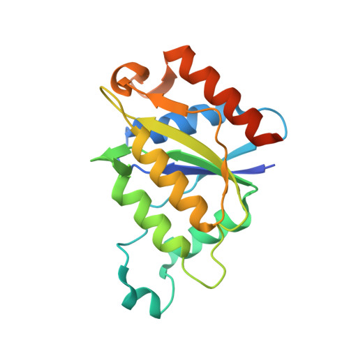
    The availability of high-intensity synchrotron facilities, technological advances in data-collection techniques and improved data-reduction and crystallographic software have ushered in a new era in high-throughput macromolecular crystallography. Here, the de novo automated crystal structure determination at 1.28 A resolution of an NAD(P)H-dependent FMN reductase flavoprotein from Pseudomonas aeruginosa PA01-derived protein Q9I4D4 using the anomalous signal from an unusually small number of S atoms is reported. Although this protein lacks the flavodoxin key fingerprint motif [(T/S)XTGXT], it has been confirmed to bind flavin mononucleotide and the binding site was identified via X-ray crystallography. This protein contains a novel flavin mononucleotide-binding site GSLRSGSYN, which has not been previously reported. Detailed statistics pertaining to sulfur phasing and other factors contributing to structure determination are discussed. Structural comparisons of the apoenzyme and the protein complexed with flavin mononucleotide show conformational changes on cofactor binding. NADPH-dependent activity has been confirmed with biochemical assays.


  • Organizational Affiliation

    Biology Department, Brookhaven National Laboratory, Upton, NY 11973, USA.


Macromolecules
Find similar proteins by:  (by identity cutoff)  |  3D Structure
Entity ID: 1
MoleculeChains Sequence LengthOrganismDetailsImage
conserved hypothetical protein
A, B
193Pseudomonas aeruginosa PAO1Mutation(s): 0 
UniProt
Find proteins for Q9I4D4 (Pseudomonas aeruginosa (strain ATCC 15692 / DSM 22644 / CIP 104116 / JCM 14847 / LMG 12228 / 1C / PRS 101 / PAO1))
Explore Q9I4D4 
Go to UniProtKB:  Q9I4D4
Entity Groups  
Sequence Clusters30% Identity50% Identity70% Identity90% Identity95% Identity100% Identity
UniProt GroupQ9I4D4
Sequence Annotations
Expand
  • Reference Sequence
Small Molecules
Experimental Data & Validation

Experimental Data

  • Method: X-RAY DIFFRACTION
  • Resolution: 2.70 Å
  • R-Value Free: 0.282 
  • R-Value Work: 0.219 
  • R-Value Observed: 0.219 
  • Space Group: P 21 21 21
Unit Cell:
Length ( Å )Angle ( ˚ )
a = 61.473α = 90
b = 65.704β = 90
c = 117.8γ = 90
Software Package:
Software NamePurpose
CNSrefinement
CBASSdata collection
HKL-2000data scaling
MOLREPphasing

Structure Validation

View Full Validation Report


Ligand Structure Quality Assessment 


Entry History 

Deposition Data

Revision History  (Full details and data files)

  • Version 1.0: 2004-08-24
    Type: Initial release
  • Version 1.1: 2008-04-30
    Changes: Version format compliance
  • Version 1.2: 2011-07-13
    Changes: Source and taxonomy, Version format compliance
  • Version 1.3: 2016-12-14
    Changes: Structure summary
  • Version 1.4: 2021-02-03
    Changes: Database references, Derived calculations, Structure summary
  • Version 1.5: 2023-08-23
    Changes: Data collection, Database references, Refinement description