1WC7

FAB FRAGMENT OF PLP-DEPENDENT CATALYTIC ANTIBODY 15A9 IN COMPLEX WITH PHOSPHOPYRIDOXYL-L-ALANINE


Experimental Data Snapshot

  • Method: X-RAY DIFFRACTION
  • Resolution: 2.33 Å
  • R-Value Free: 0.309 
  • R-Value Observed: 0.222 

wwPDB Validation   3D Report Full Report


Ligand Structure Quality Assessment 


This is version 1.4 of the entry. See complete history


Literature

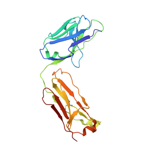
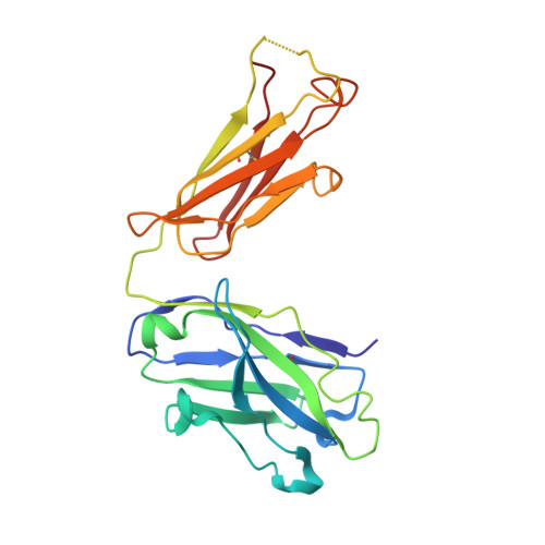
Structure of a Pseudomerohedrally Twinned Monoclinic Crystal Form of a Pyridoxal Phosphate-Dependent Catalytic Antibody

Golinelli-Pimpaneau, B.

(2005) Acta Crystallogr D Biol Crystallogr 61: 472

  • DOI: https://doi.org/10.1107/S0907444905003331
  • Primary Citation of Related Structures:  
    1WC7

  • PubMed Abstract: 

    The purification, crystallization and structure determination at 2.3 A resolution of the complex of the pyridoxal-5'-phosphate (PLP) dependent catalytic antibody 15A9 with a phosphopyridoxyl-L-alanine (PPL-L-alanine) substrate analogue are described. The crystal belongs to space group P2(1), with two molecules in the asymmetric unit related by non-crystallographic symmetry. The unit-cell parameters are a = 63.5, b = 81.7, c = 79.3 A and beta is fortuitously 90 degrees . Refinement of the structure converged at unacceptably high R factors. Although the traditional analysis of intensity distribution did not indicate twinning, pseudomerohedral twinning was revealed by a newer test based on local intensity differences [Padilla & Yeates (2003), Acta Cryst. D59, 1124-1130]. When the potential twinning operator was included in SHELX, the structure could be satisfactorily refined with a twinning fraction of 0.46, indicating a nearly perfect hemihedrally twinned crystal. One of the active sites is occupied by the phosphopyridoxyl-L-alanine ligand, while one iodide ion mimics the cofactor phosphate group in the other. Four other iodide ions are present in the structure: two are involved in specific intermolecular contacts and two dictate the conformation of the CDRH3 loop in each molecule.


  • Organizational Affiliation

    Laboratoire d'Enzymologie et de Biochimie Structurales, Bâtiment 34, CNRS, 1 Avenue de la Terrasse, 91190 Gif-sur-Yvette, France. beatrice.golinelli@lebs.cnrs-gif.fr


Macromolecules
Find similar proteins by:  (by identity cutoff)  |  3D Structure
Entity ID: 1
MoleculeChains Sequence LengthOrganismDetailsImage
FAB FRAGMENT OF CATALYTIC ANTIBODY 15A9, LIGHT CHAINA,
D [auth L]
213Mus musculusMutation(s): 0 
Entity Groups  
Sequence Clusters30% Identity50% Identity70% Identity90% Identity95% Identity100% Identity
Sequence Annotations
Expand
  • Reference Sequence
Find similar proteins by:  (by identity cutoff)  |  3D Structure
Entity ID: 2
MoleculeChains Sequence LengthOrganismDetailsImage
FAB FRAGMENT OF CATALYTIC ANTIBODY 15A9, HEAVY CHAINB,
C [auth H]
226Mus musculusMutation(s): 0 
Entity Groups  
Sequence Clusters30% Identity50% Identity70% Identity90% Identity95% Identity100% Identity
Sequence Annotations
Expand
  • Reference Sequence
Experimental Data & Validation

Experimental Data

  • Method: X-RAY DIFFRACTION
  • Resolution: 2.33 Å
  • R-Value Free: 0.309 
  • R-Value Observed: 0.222 
  • Space Group: P 1 21 1
Unit Cell:
Length ( Å )Angle ( ˚ )
a = 63.476α = 90
b = 81.691β = 89.98
c = 79.33γ = 90
Software Package:
Software NamePurpose
SHELXL-97refinement
DENZOdata reduction
SCALEPACKdata scaling
AMoREphasing

Structure Validation

View Full Validation Report


Ligand Structure Quality Assessment 


Entry History 

Deposition Data

Revision History  (Full details and data files)

  • Version 1.0: 2005-11-30
    Type: Initial release
  • Version 1.1: 2011-05-08
    Changes: Version format compliance
  • Version 1.2: 2011-07-13
    Changes: Version format compliance
  • Version 1.3: 2019-01-30
    Changes: Data collection, Experimental preparation, Other
  • Version 1.4: 2019-05-22
    Changes: Data collection, Refinement description