1UWI

CRYSTAL STRUCTURE OF MUTATED BETA-GLYCOSIDASE FROM SULFOLOBUS SOLFATARICUS, WORKING AT MODERATE TEMPERATURE


Experimental Data Snapshot

  • Method: X-RAY DIFFRACTION
  • Resolution: 2.55 Å
  • R-Value Free: 0.241 
  • R-Value Work: 0.209 
  • R-Value Observed: 0.209 

wwPDB Validation   3D Report Full Report


This is version 1.2 of the entry. See complete history


Literature

Comparative Study and Mutational Analysis of Distinctive Structural Elements of Hyperthermophilic Enzymes.

Leon, M.Isorna, P.Menendez, M.Sanz-Aparicio, J.Polaina, J.

(2007) Protein J 26: 435

  • DOI: https://doi.org/10.1007/s10930-007-9083-2
  • Primary Citation of Related Structures:  
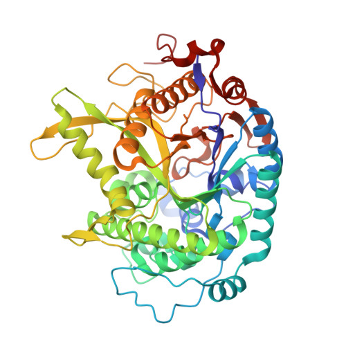
    1UWI

  • PubMed Abstract: 

    Comparison of the three-dimensional structure of hyperthermophilic and mesophilic beta-glycosidases shows differences in secondary structure composition. The enzymes from hyperthermophilic archaea have a significantly larger number of beta-strands arranged in supernumerary beta-sheets compared to mesophilic enzymes from bacteria and other organisms. Amino acid replacements designed to alter the structure of the supernumerary beta-strands were introduced by site directed mutagenesis into the sequence encoding the beta-glycosidase from Sulfolobus solfataricus. Most of the replacements caused almost complete loss of activity but some yielded enzyme variants whose activities were affected specifically at higher temperatures. Far-UV CD spectra recorded as a function of temperature for both wild type beta-glycosidase and mutant V349G, one of the mutants with reduced activity at higher temperatures, were similar, showing that the protein structure of the mutant was stable at the highest temperatures assayed. The properties of mutant V349G show a difference between thermostability (stability of the protein structure at high temperatures) and thermophilicity (optimal activity at high temperatures).


  • Organizational Affiliation

    Instituto de Agroquímica y Tecnología de Alimentos, Consejo Superior de Investigaciones Científicas, Apdo. de Correos 73, Burjassot, Valencia, E46100, Spain.


Macromolecules
Find similar proteins by:  (by identity cutoff)  |  3D Structure
Entity ID: 1
MoleculeChains Sequence LengthOrganismDetailsImage
BETA-GALACTOSIDASE
A, B, C, D
489Saccharolobus solfataricus P2Mutation(s): 4 
EC: 3.2.1.23
UniProt
Find proteins for P22498 (Saccharolobus solfataricus (strain ATCC 35092 / DSM 1617 / JCM 11322 / P2))
Explore P22498 
Go to UniProtKB:  P22498
Entity Groups  
Sequence Clusters30% Identity50% Identity70% Identity90% Identity95% Identity100% Identity
UniProt GroupP22498
Sequence Annotations
Expand
  • Reference Sequence
Experimental Data & Validation

Experimental Data

  • Method: X-RAY DIFFRACTION
  • Resolution: 2.55 Å
  • R-Value Free: 0.241 
  • R-Value Work: 0.209 
  • R-Value Observed: 0.209 
  • Space Group: P 21 21 21
Unit Cell:
Length ( Å )Angle ( ˚ )
a = 79.082α = 90
b = 132.425β = 90
c = 219.149γ = 90
Software Package:
Software NamePurpose
CNSrefinement
MOSFLMdata reduction
SCALEPACKdata scaling
AMoREphasing

Structure Validation

View Full Validation Report


Entry History 

Deposition Data

Revision History  (Full details and data files)

  • Version 1.0: 2005-02-09
    Type: Initial release
  • Version 1.1: 2014-07-30
    Changes: Data collection, Database references, Derived calculations, Non-polymer description, Other, Refinement description, Source and taxonomy, Structure summary, Version format compliance
  • Version 1.2: 2023-12-13
    Changes: Data collection, Database references, Other, Refinement description