1US4

PUTATIVE GLUR0 LIGAND BINDING CORE WITH L-GLUTAMATE


Experimental Data Snapshot

  • Method: X-RAY DIFFRACTION
  • Resolution: 1.75 Å
  • R-Value Free: 0.206 
  • R-Value Work: 0.178 
  • R-Value Observed: 0.178 

wwPDB Validation   3D Report Full Report


This is version 1.5 of the entry. See complete history


Literature

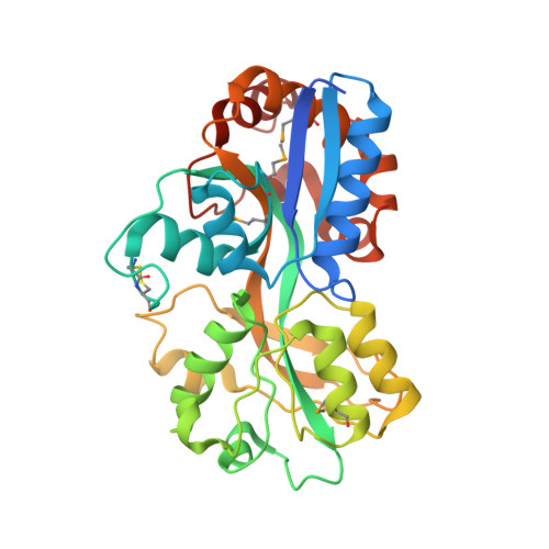
Structure of the Thermus Thermophilus Putative Periplasmic Glutamate/Glutamine-Binding Protein

Takahashi, H.Inagaki, E.Kuroishi, C.Tahirov, T.H.

(2004) Acta Crystallogr D Biol Crystallogr 60: 1846

  • DOI: https://doi.org/10.1107/S0907444904019420
  • Primary Citation of Related Structures:  
    1US4, 1US5

  • PubMed Abstract: 

    As part of a structural genomics project, the crystal structure of a 314-amino-acid protein encoded by Thermus thermophilus HB8 gene TT1099 was solved to 1.75 A using the multiple-wavelength anomalous dispersion (MAD) method and a selenomethionine-incorporated protein. The native protein structure was solved to 1.5 A using the molecular-replacement method. Both structures revealed a bound ligand, L-glutamate or L-glutamine, and a fold related to the periplasmic substrate-binding proteins (PSBP). Further comparative structural analysis with other PSBP-fold proteins revealed the conservation of the predicted membrane permease binding surface area and indicated that the T. thermophilus HB8 molecule is most likely to be an L-glutamate and/or an L-glutamine-binding protein related to the cluster 3 periplasmic receptors. However, the geometry of ligand binding is unique to the T. thermophilus HB8 molecule.


  • Organizational Affiliation

    Highthroughput Factory, RIKEN Harima Institute, 1-1-1 Kouto, Mikazuki-cho, Sayo-gun, Hyogo 679-5148, Japan.


Macromolecules
Find similar proteins by:  (by identity cutoff)  |  3D Structure
Entity ID: 1
MoleculeChains Sequence LengthOrganismDetailsImage
PUTATIVE GLUR0 LIGAND BINDING CORE314Thermus thermophilus HB8Mutation(s): 0 
UniProt
Find proteins for P83817 (Thermus thermophilus)
Explore P83817 
Go to UniProtKB:  P83817
Entity Groups  
Sequence Clusters30% Identity50% Identity70% Identity90% Identity95% Identity100% Identity
UniProt GroupP83817
Sequence Annotations
Expand
  • Reference Sequence
Small Molecules
Modified Residues  1 Unique
IDChains TypeFormula2D DiagramParent
MSE
Query on MSE
A
L-PEPTIDE LINKINGC5 H11 N O2 SeMET
Experimental Data & Validation

Experimental Data

  • Method: X-RAY DIFFRACTION
  • Resolution: 1.75 Å
  • R-Value Free: 0.206 
  • R-Value Work: 0.178 
  • R-Value Observed: 0.178 
  • Space Group: P 21 21 21
Unit Cell:
Length ( Å )Angle ( ˚ )
a = 51.518α = 90
b = 68.092β = 90
c = 81.381γ = 90
Software Package:
Software NamePurpose
HKL-2000data reduction
HKL-2000data scaling
CNSphasing
ARP/wARPphasing
CNSrefinement

Structure Validation

View Full Validation Report


Entry History 

Deposition Data

Revision History  (Full details and data files)

  • Version 1.0: 2003-11-19
    Type: Initial release
  • Version 1.1: 2011-05-08
    Changes: Version format compliance
  • Version 1.2: 2011-07-13
    Changes: Version format compliance
  • Version 1.3: 2017-06-28
    Changes: Refinement description
  • Version 1.4: 2019-03-06
    Changes: Data collection, Derived calculations, Experimental preparation
  • Version 1.5: 2019-05-22
    Changes: Data collection, Experimental preparation