1SCF

HUMAN RECOMBINANT STEM CELL FACTOR


Experimental Data Snapshot

  • Method: X-RAY DIFFRACTION
  • Resolution: 2.20 Å
  • R-Value Free: 0.242 
  • R-Value Work: 0.199 
  • R-Value Observed: 0.199 

wwPDB Validation   3D Report Full Report


This is version 1.3 of the entry. See complete history


Literature

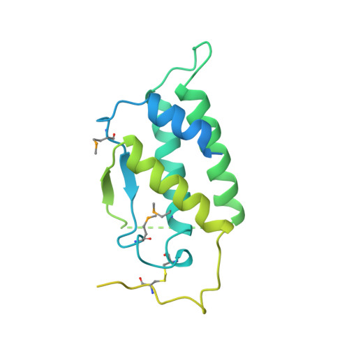
Structure of the active core of human stem cell factor and analysis of binding to its receptor kit.

Jiang, X.Gurel, O.Mendiaz, E.A.Stearns, G.W.Clogston, C.L.Lu, H.S.Osslund, T.D.Syed, R.S.Langley, K.E.Hendrickson, W.A.

(2000) EMBO J 19: 3192-3203

  • DOI: https://doi.org/10.1093/emboj/19.13.3192
  • Primary Citation of Related Structures:  
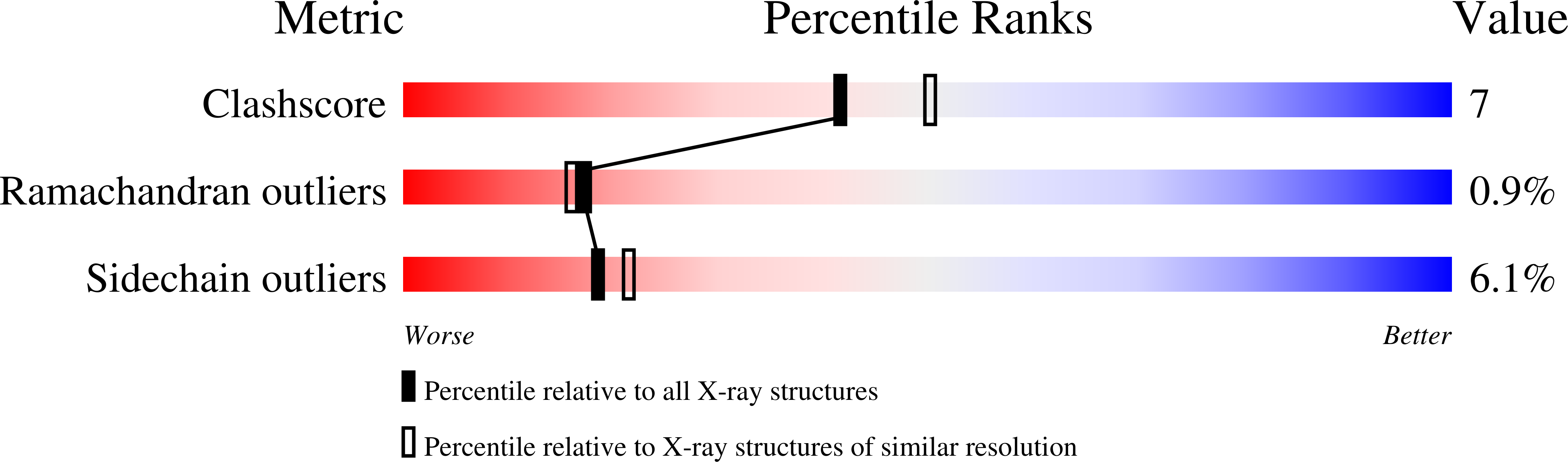
    1SCF

  • PubMed Abstract: 

    Stem cell factor (SCF) is an early-acting hematopoietic cytokine that elicits multiple biological effects. SCF is dimeric and occurs in soluble and membrane-bound forms. It transduces signals by ligand- mediated dimerization of its receptor, Kit, which is a receptor tyrosine kinase related to the receptors for platelet-derived growth factor (PDGF), macrophage colony-stimulating factor, Flt-3 ligand and vascular endothelial growth factor (VEGF). All of these have extracellular ligand-binding portions composed of immunoglobulin-like repeats. We have determined the crystal structure of selenomethionyl soluble human SCF at 2.2 A resolution by multiwavelength anomalous diffraction phasing. SCF has the characteristic helical cytokine topology, but the structure is unique apart from core portions. The SCF dimer has a symmetric 'head-to-head' association. Using various prior observations, we have located potential Kit-binding sites on the SCF dimer. A superimposition of this dimer onto VEGF in its complex with the receptor Flt-1 places the binding sites on SCF in positions of topographical and electrostatic complementarity with the Kit counterparts of Flt-1, and a similar model can be made for the complex of PDGF with its receptor.


  • Organizational Affiliation

    Department of Biochemistry and Molecular Biophysics and Howard Hughes Medical Institute, Columbia University, New York, NY 10032, USA.


Macromolecules
Find similar proteins by:  (by identity cutoff)  |  3D Structure
Entity ID: 1
MoleculeChains Sequence LengthOrganismDetailsImage
STEM CELL FACTOR
A, B, C, D
273Homo sapiensMutation(s): 3 
UniProt & NIH Common Fund Data Resources
Find proteins for P21583 (Homo sapiens)
Explore P21583 
Go to UniProtKB:  P21583
PHAROS:  P21583
GTEx:  ENSG00000049130 
Entity Groups  
Sequence Clusters30% Identity50% Identity70% Identity90% Identity95% Identity100% Identity
UniProt GroupP21583
Sequence Annotations
Expand
  • Reference Sequence
Small Molecules
Modified Residues  1 Unique
IDChains TypeFormula2D DiagramParent
MSE
Query on MSE
A, B, C, D
L-PEPTIDE LINKINGC5 H11 N O2 SeMET
Experimental Data & Validation

Experimental Data

  • Method: X-RAY DIFFRACTION
  • Resolution: 2.20 Å
  • R-Value Free: 0.242 
  • R-Value Work: 0.199 
  • R-Value Observed: 0.199 
  • Space Group: P 21 21 21
Unit Cell:
Length ( Å )Angle ( ˚ )
a = 71.82α = 90
b = 82.55β = 90
c = 88.19γ = 90
Software Package:
Software NamePurpose
DENZOdata reduction
SCALEPACKdata scaling
MADLSQmodel building
X-PLORmodel building
X-PLORrefinement
MADLSQphasing
X-PLORphasing

Structure Validation

View Full Validation Report


Entry History 

Deposition Data

Revision History  (Full details and data files)

  • Version 1.0: 2000-07-07
    Type: Initial release
  • Version 1.1: 2008-03-24
    Changes: Version format compliance
  • Version 1.2: 2011-07-13
    Changes: Version format compliance
  • Version 1.3: 2022-12-21
    Changes: Database references, Derived calculations