1O2G

Elaborate Manifold of Short Hydrogen Bond Arrays Mediating Binding of Active Site-Directed Serine Protease Inhibitors


Experimental Data Snapshot

  • Method: X-RAY DIFFRACTION
  • Resolution: 1.58 Å
  • R-Value Free: 0.250 
  • R-Value Work: 0.219 
  • R-Value Observed: 0.222 

wwPDB Validation   3D Report Full Report


Ligand Structure Quality Assessment 


This is version 2.0 of the entry. See complete history


Literature

Elaborate Manifold of Short Hydrogen Bond Arrays Mediating Binding of Active Site-Directed Serine Protease Inhibitors

Katz, B.A.Elrod, K.Verner, E.Mackman, R.L.Luong, C.Shrader, W.D.Sendzik, M.Spencer, J.R.Sprengeler, P.A.Kolesnikov, A.Tai, V.W.-F.Hui, H.C.Breitenbucher, J.G.Allen, D.Janc, J.W.

(2003) J Mol Biol 329: 93-120

  • DOI: https://doi.org/10.1016/s0022-2836(03)00399-1
  • Primary Citation of Related Structures:  
    1O2G, 1O2H, 1O2I, 1O2J, 1O2K, 1O2L, 1O2M, 1O2N, 1O2O, 1O2P, 1O2Q, 1O2R, 1O2S, 1O2T, 1O2U, 1O2V, 1O2W, 1O2X, 1O2Y, 1O2Z, 1O30, 1O31, 1O32, 1O33, 1O34, 1O35, 1O36, 1O37, 1O38, 1O39, 1O3A, 1O3B, 1O3C, 1O3D, 1O3E, 1O3F, 1O3G, 1O3H, 1O3I, 1O3J, 1O3K, 1O3L, 1O3M, 1O3N, 1O3O, 1O3P

  • PubMed Abstract: 

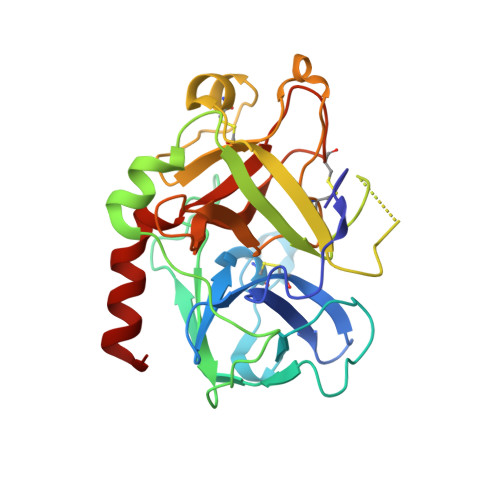
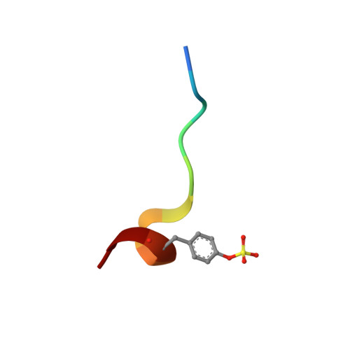
    An extensive structural manifold of short hydrogen bond-mediated, active site-directed, serine protease inhibition motifs is revealed in a set of over 300 crystal structures involving a large suite of small molecule inhibitors (2-(2-phenol)-indoles and 2-(2-phenol)-benzimidazoles) determined over a wide range of pH (3.5-11.4). The active site hydrogen-bonding mode was found to vary markedly with pH, with the steric and electronic properties of the inhibitor, and with the type of protease (trypsin, thrombin or urokinase type plasminogen activator (uPA)). The pH dependence of the active site hydrogen-bonding motif is often intricate, constituting a distinct fingerprint of each complex. Isosteric replacements or minor substitutions within the inhibitor that modulate the pK(a) of the phenol hydroxyl involved in short hydrogen bonding, or that affect steric interactions distal to the active site, can significantly shift the pH-dependent structural profile characteristic of the parent scaffold, or produce active site-binding motifs unique to the bound analog. Ionization equilibria at the active site associated with inhibitor binding are probed in a series of the protease-inhibitor complexes through analysis of the pH dependence of the structure and environment of the active site-binding groups involved in short hydrogen bond arrays. Structures determined at high pH (>11), suggest that the pK(a) of His57 is dramatically elevated, to a value as high as approximately 11 in certain complexes. K(i) values involving uPA and trypsin determined as a function of pH for a set of inhibitors show pronounced parabolic pH dependence, the pH for optimal inhibition governed by the pK(a) of the inhibitor phenol involved in short hydrogen bonds. Comparison of structures of trypsin, thrombin and uPA, each bound by the same inhibitor, highlights important structural variations in the S1 and active sites accessible for engineering notable selectivity into remarkably small molecules with low nanomolar K(i) values.


  • Organizational Affiliation

    Celera, 180 Kimball Way, South San Francisco, CA 94080, USA. brad.katz@celera.com


Macromolecules
Find similar proteins by:  (by identity cutoff)  |  3D Structure
Entity ID: 1
MoleculeChains Sequence LengthOrganismDetailsImage
THROMBINA [auth L]36Homo sapiensMutation(s): 0 
EC: 3.4.21.5
UniProt & NIH Common Fund Data Resources
Find proteins for P00734 (Homo sapiens)
Explore P00734 
Go to UniProtKB:  P00734
PHAROS:  P00734
GTEx:  ENSG00000180210 
Entity Groups  
Sequence Clusters30% Identity50% Identity70% Identity90% Identity95% Identity100% Identity
UniProt GroupP00734
Sequence Annotations
Expand
  • Reference Sequence
Find similar proteins by:  (by identity cutoff)  |  3D Structure
Entity ID: 2
MoleculeChains Sequence LengthOrganismDetailsImage
THROMBINB [auth H]259Homo sapiensMutation(s): 0 
EC: 3.4.21.5
UniProt & NIH Common Fund Data Resources
Find proteins for P00734 (Homo sapiens)
Explore P00734 
Go to UniProtKB:  P00734
PHAROS:  P00734
GTEx:  ENSG00000180210 
Entity Groups  
Sequence Clusters30% Identity50% Identity70% Identity90% Identity95% Identity100% Identity
UniProt GroupP00734
Sequence Annotations
Expand
  • Reference Sequence

Find similar proteins by:  Sequence   |   3D Structure  

Entity ID: 3
MoleculeChains Sequence LengthOrganismDetailsImage
ACETYL HIRUDINC [auth I]11Hirudo medicinalisMutation(s): 0 
UniProt
Find proteins for P28504 (Hirudo medicinalis)
Explore P28504 
Go to UniProtKB:  P28504
Entity Groups  
Sequence Clusters30% Identity50% Identity70% Identity90% Identity95% Identity100% Identity
UniProt GroupP28504
Sequence Annotations
Expand
  • Reference Sequence
Small Molecules
Ligands 2 Unique
IDChains Name / Formula / InChI Key2D Diagram3D Interactions
696
Query on 696

Download Ideal Coordinates CCD File 
E [auth H]3-{5-[AMINO(IMINIO)METHYL]-1H-INDOL-2-YL}-1,1'-BIPHENYL-2-OLATE
C21 H17 N3 O
GAVRMVQHHVMXFD-UHFFFAOYSA-N
NA
Query on NA

Download Ideal Coordinates CCD File 
D [auth H]SODIUM ION
Na
FKNQFGJONOIPTF-UHFFFAOYSA-N
Modified Residues  1 Unique
IDChains TypeFormula2D DiagramParent
TYS
Query on TYS
C [auth I]L-PEPTIDE LINKINGC9 H11 N O6 STYR
Binding Affinity Annotations 
IDSourceBinding Affinity
696 BindingDB:  1O2G Ki: min: 320, max: 760 (nM) from 2 assay(s)
PDBBind:  1O2G Ki: 760 (nM) from 1 assay(s)
Binding MOAD:  1O2G Ki: 760 (nM) from 1 assay(s)
Experimental Data & Validation

Experimental Data

  • Method: X-RAY DIFFRACTION
  • Resolution: 1.58 Å
  • R-Value Free: 0.250 
  • R-Value Work: 0.219 
  • R-Value Observed: 0.222 
  • Space Group: C 1 2 1
Unit Cell:
Length ( Å )Angle ( ˚ )
a = 71.36α = 90
b = 72.06β = 101.77
c = 72.96γ = 90
Software Package:
Software NamePurpose
CrystalCleardata collection
CrystalCleardata reduction
X-PLORrefinement
CrystalCleardata scaling
Quantamodel building

Structure Validation

View Full Validation Report


Ligand Structure Quality Assessment 


Entry History 

Revision History  (Full details and data files)

  • Version 1.0: 2003-05-13
    Type: Initial release
  • Version 1.1: 2008-04-26
    Changes: Version format compliance
  • Version 1.2: 2011-07-13
    Changes: Atomic model, Database references, Derived calculations, Non-polymer description, Structure summary, Version format compliance
  • Version 1.3: 2012-12-12
    Changes: Other
  • Version 1.4: 2013-03-13
    Changes: Other
  • Version 2.0: 2023-11-15
    Changes: Atomic model, Data collection, Database references, Derived calculations